Abstract
Background: Beginning in 2020–2021, embalmers in multiple countries reported observing large, tough, rubbery white or off-white fibrous structures in the veins and arteries of embalmed corpses, which they described as distinct from classic postmortem clots. Methods: We conducted four annual cross-sectional surveys (2022–2025) of active embalmers in the United States, Canada, United Kingdom, Australia, and New Zealand using SurveyMonkey. A dual distribution strategy (professional associations and direct emails to funeral homes) was used. Core questions assessed observation of unusual white fibrous structures and estimated percentage of corpses affected. Results: Across 808 total responses, the proportion of embalmers reporting observation of these structures ranged from 66% to 83%. Weighted average prevalence in affected corpses ranged from 19% to 27%. The 2022 survey showed a marked increase in first observations beginning in 2020 and accelerating in 2021. Conclusions: Multiple years of surveys document consistent self-reported observations by experienced embalmers of unusual white fibrous structures in a substantial fraction of corpses, with a clear increase noted around 2020–2021. These findings constitute a potential safety signal that warrants independent investigation by forensic pathologists and biomedical researchers to characterize the structures and determine their etiology.
Keywords
Embalmers Postmortem clots White fibrous clots Survey Forensic pathology Observational study.
Introduction
In forensic pathology and thanatology, postmortem blood clots (cruors) are well-described. Classic types include “chicken-fat” clots (yellowish, friable plasma layer) and “currant-jelly” clots (dark red, RBC-rich). These are typically non-adherent to vessel walls and differ from antemortem thrombi in consistency and appearance [1,2].
Beginning in late 2021 and into 2022, embalmers and funeral directors in multiple countries began reporting observations of large, tough, rubbery, white or off-white fibrous structures in the veins and arteries of embalmed corpses. Experienced practitioners described these as unlike any postmortem clots encountered in decades of practice, larger, more extensive, tougher, and different in color and handling from traditional chicken-fat or currant-jelly clots.
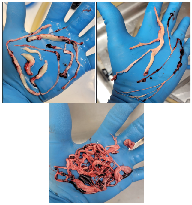
Experienced embalmers have consistently described these structures using specific operational criteria that distinguish them from classic postmortem clots. The structures are typically large (often several inches to over a foot in length, occasionally approaching the length of a limb), tough and rubbery in texture (elastic, resistant to tearing or fragmentation, and sometimes compared to calamari or earthworms), predominantly white or off-white, and extensively fill or conform to the vascular lumen. Many embalmers report that these formations impede normal drainage of blood and hinder uniform distribution of embalming fluid, frequently requiring additional injection sites, higher pressures, or specialized techniques to achieve adequate perfusion, phenomena they describe as more frequent and more severe than with traditional postmortem clots. Photographic documentation provided by multiple practitioners (e.g., Figure 1) illustrates the gross appearance of long, cohesive, fibrous casts that embalmers state differ markedly in handling and extent from the softer, more friable, or layered classic chicken-fat and currant-jelly clots.
The embalming process itself involves arterial injection of formaldehyde-based preservative fluids under pressure, typically preceded by drainage of blood. Formaldehyde rapidly cross-links and coagulates proteins, which can fix existing clots in place, alter their texture and color, and modify their handling characteristics. Embalmers routinely employ water conditioners and anticoagulants in the fluid to minimize additional clotting during the procedure. Despite these standard practices, and the fact that experienced practitioners account for typical postmortem and iatrogenic changes, many report that the unusual white fibrous structures remain distinctly more obstructive, rubbery, and extensive than structures encountered in prior decades. This does not prove the structures predate embalming or are entirely independent of it; rather, it underscores that definitive characterization requires laboratory methods beyond gross observation during routine embalming.
Early public reports came from UK funeral director John O’Looney and Alabama embalmer Richard Hirschman, the latter providing photographic documentation, see figure 1. Similar accounts subsequently emerged from other embalmers, including presentations at professional meetings. In October 2022, Indiana embalmer Wallace Hooker reported that a majority of approximately 100 embalmers attending an Ohio Embalmers Association (OEA) convention had confirmed observing such structures over the preceding 16–18 months.
Following these reports, the lead author contacted OEA leadership in November 2022. OEA Vice President Woody Wilson confirmed having personally observed the structures during embalming at his funeral home in Marysville, Ohio, and voluntarily provided photographic examples via email in January 2023. This independent professional confirmation from an officer of a state embalmer association was a key factor in the decision to conduct systematic multi-year surveys to quantify the prevalence and timing of these observations.
The objective of this study was to document, through anonymous surveys, the prevalence of reported observations of these unusual white fibrous structures, the proportion of corpses affected, and the temporal pattern of first appearance.
Materials and Methods
This study consisted of four annual cross-sectional online surveys (2022–2025) administered through SurveyMonkey. The target population was active embalmers performing embalming in the United States, Canada, United Kingdom, Australia, and New Zealand.
A dual distribution strategy was used. In the top-down approach, survey links were emailed to approximately 50 national, regional, and state funeral director and embalmer associations, with requests to forward to members. Telephone follow-up was conducted with U.S. and Canadian associations. In the bottom-up approach, direct emails were sent to over 1,700 funeral homes in major cities across the five countries.
The 2022 survey collected data on observation of large white fibrous structures, estimated percentage of corpses affected, and year of first observation. Subsequent surveys retained core questions on observation and prevalence, with additional items on micro-clotting and age stratification in later waves. The “year first observed” question was removed after 2022 to reduce recall burden.
Participation was voluntary and anonymous. As an anonymous survey of professionals regarding their observations, the study was determined exempt from institutional review board oversight. Data were analyzed descriptively. Weighted averages for percentage of corpses affected included 0% responses from embalmers who reported never observing the structures.
Ethical Statement
This study involved an anonymous, cross-sectional online survey of active deathcare professionals regarding aggregate, non-identifiable workplace observations. No private, identifiable personal or medical data was collected from the participants or the deceased individuals referenced. Because the research involves only voluntary, completely anonymous surveys of professionals regarding aggregate business observations, with no direct intervention, clinical interaction, or access to private patient records, the study satisfies the criteria for exemption under standard institutional review board (IRB) and human subjects protection regulations (e.g., 45 CFR 46.104).
Results
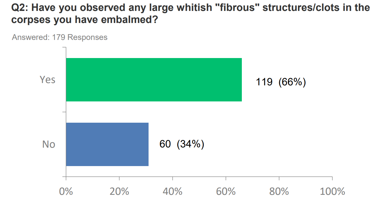
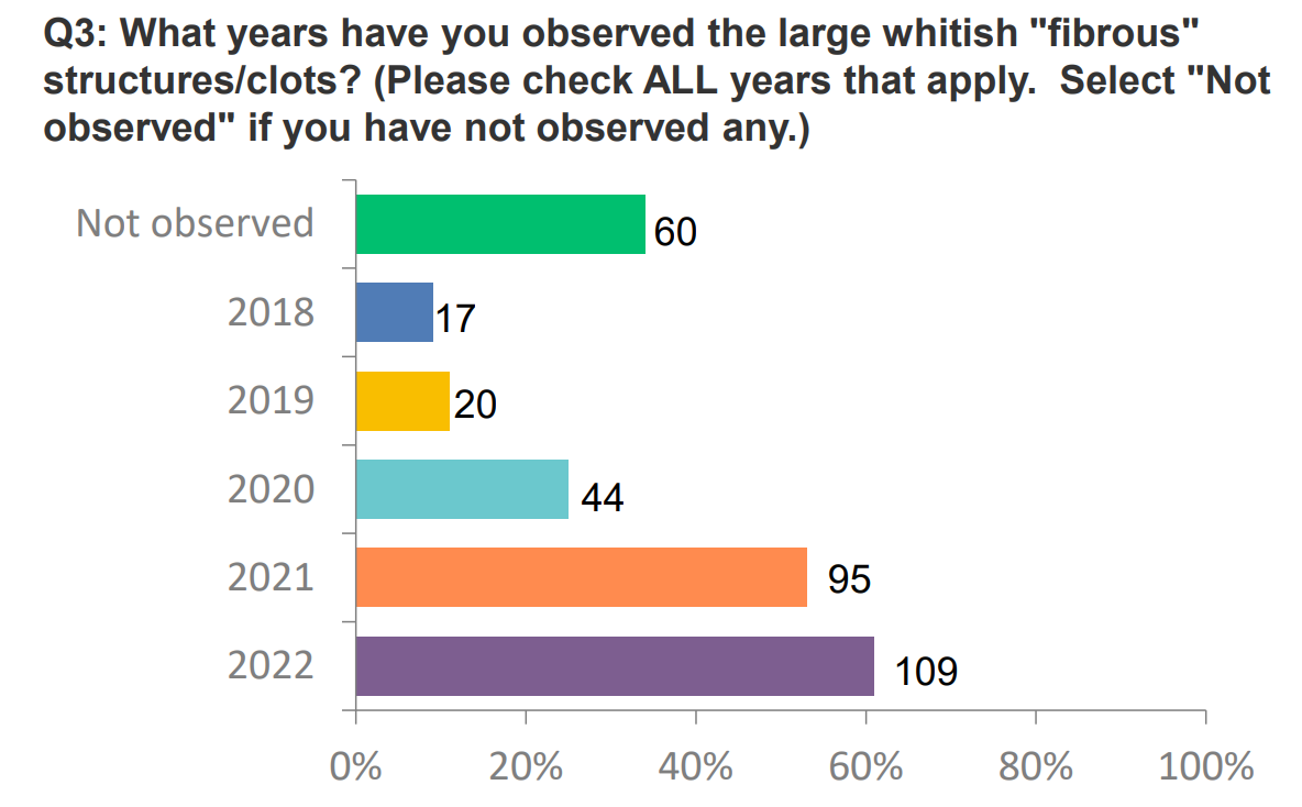
A total of 808 embalmers responded across the four survey years (Table 1). In the 2022 survey, 66% of respondents reported having observed the unusual white fibrous structures (Figure 2). Among those who had observed them, only 14–17% reported seeing them in 2018 or 2019, increasing to 37% for 2020 and 80% for 2021 (Figure 3), indicating a clear temporal rise in first observations.
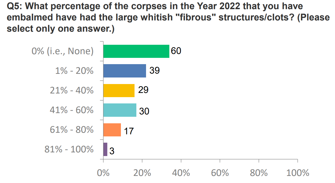
Respondents also estimated the proportion of corpses in 2022 that contained these structures. Reported values varied widely, with the largest group indicating 0%, but substantial numbers reporting 1–20%, 21–40%, 41–60%, and smaller proportions reporting 61–80% or 81–100% (Figure 4). These distributions contributed to a weighted average prevalence of 24% for 2022.
| Survey Year | Embalmers (n) | Reporting Structures n (%) | Weighted Avg. % Corpses |
| 2022 | 179 | 119 (66%) | 24% |
| 2023 | 269 | 197 (73%) | 20% |
| 2024 | 301 | 250 (83%) | 27% |
| 2025 | 59 | 42 (71%) | 19% |
Note: The 2025 survey had a substantially smaller sample size, which reduces precision.
Discussion and Limitations
Over four annual surveys, several hundred embalmers across five countries consistently reported observing unusual white fibrous structures in embalmed corpses. The proportion reporting these observations remained high (66–83%), and the estimated prevalence in affected cases was substantial (19–27%; Figure 4 shows the 2022 distribution). The 2022 data demonstrated a temporal increase in first observations beginning in 2020 and rising further in 2021. These self-reported findings align with earlier anecdotal accounts from experienced embalmers and were described using consistent operational criteria, including large size, rubbery or tough elastic texture, extensive filling of the vascular lumen, and interference with embalming fluid distribution.
These findings constitute a legitimate frontline signal, but several limitations must be considered. Participation was voluntary and relied on non-probability sampling through professional associations and direct emails. Embalmers who had noticed unusual findings may have been more likely to respond, introducing selection and response bias. True population response rates cannot be determined. All data are self-reported; no standardized definitions or photographic, histological or biochemical confirmation was required. Although experienced embalmers are skilled observers, inter-observer variability and potential confirmation bias following media coverage cannot be excluded. The question on year of first observation (asked only in 2022) is subject to recall bias. The 2025 sample size was small (n=59), reducing precision.
While embalmers consistently described the structures as distinct from classic postmortem clots using the operational criteria noted above, definitive differentiation from known postmortem phenomena, agonal thrombi, COVID-associated fibrin alterations, or embalming-related modifications requires laboratory methods (histopathology, immunohistochemistry, proteomics, vibrational spectroscopy/Raman analysis, or electron microscopy) [3,4]. Such analyses are essential to determine composition, ultrastructure, and any relationship to amyloidogenic fibrin aggregates or other entities.
Alternative explanations for the temporal pattern warrant careful consideration. SARS-CoV-2 infection is associated with hypercoagulability and increased thrombotic events [5]. Pandemic-related factors, including excess mortality, changes in postmortem intervals, and shifts in death certification practices, may have altered the cases encountered by embalmers. Increased awareness following media coverage beginning in late 2022 may also have heightened vigilance regarding vascular findings. The embalming process itself (formaldehyde cross-linking, fluid dynamics, and use of conditioners) can modify the gross appearance of intravascular material; experienced practitioners account for these effects, yet still report the structures as distinct.
Despite these limitations, the repeated and internally consistent reports from frontline professionals who routinely examine vascular contents during embalming represent a credible observational signal worthy of independent scientific investigation. The value of this study lies in systematically documenting these reports and identifying a phenomenon that requires rigorous laboratory followup.
Conclusions
Multi-year surveys of embalmers in five countries document consistent self-reported observations of unusual white fibrous structures in a substantial fraction of embalmed corpses, with a marked increase noted around 2020–2021. While causation cannot be inferred, these findings constitute a potential safety signal that warrants prompt, independent investigation. Priority should be given to histopathological, biochemical, proteomic, and spectroscopic characterization of these structures to determine their composition, distinguish them rigorously from classic postmortem clots and other known entities, and clarify any implications for living patients. Frontline observations by experienced embalmers provide valuable sentinel information that can guide targeted scientific inquiry, but only objective laboratory investigation can establish whether these represent a novel pathological process or variants of recognized phenomena amplified by pandemic-era factors.
Declarations
Author Contributions
T.F.H.: Conceptualization, survey design and distribution, data collection and analysis, manuscript drafting and revision. L.K.: Survey methodology, SurveyMonkey implementation, data management, manuscript review. D.S.: Manuscript revision, scientific framing, and preparation for preprint submission. All authors read and approved the final manuscript.
Funding
This study and all surveys were funded entirely by the T.F.H. and L.K. No external funding or grants were received.
Data Availability
De-identified aggregate survey data and raw SurveyMonkey response files are available from the corresponding author upon reasonable request, subject to privacy considerations.
Conflicts of Interest
The authors declare that they have no competing interests or financial ties relevant to this study.
Acknowledgments
We thank the embalmers and funeral directors who participated in the surveys and the professional associations that assisted with distribution. We also acknowledge early confirmations and photographic documentation provided by members of the Ohio Embalmers Association.
AI disclosure
The author used artificial intelligence, assisted tools solely for reference formatting and citation verification. All original ideas, analyses, interpretations, and textual content were conceived and written by the author.
References
- Solarino, B., Ambrosi, L., Benevento, M., Ferorelli, D., Buschmann, C., & Nicolì, S. (2025). Cadaver clots: a systematic review of the literature. Forensic science, medicine, and pathology, 21(4), 1831–1842. DOI ↗ Google Scholar ↗
- Rizac, R. I., Tiu, R. E., Turbatu, R. M., & Ciobotaru-Pîrvu, E. (2021). Thrombi, post-mortem clots and agonal thrombi: How to tell the difference? Revista Română de Medicină Veterinară, 31(4), 36–40. DOI ↗ Google Scholar ↗
- Kruger, A., Vlok, M., Turner, S., Venter, C., Laubscher, G. J., Kell, D. B., & Pretorius, E. (2022). Proteomics of fibrin amyloid microclots in long COVID/post-acute sequelae of COVID-19 (PASC) shows many entrapped pro-inflammatory molecules that may also contribute to a failed fibrinolytic system. Cardiovascular diabetology, 21(1), 190. DOI ↗ Google Scholar ↗
- Westman, H., Hammarström, P., & Nyström, S. (2025). SARS-CoV-2 Spike Protein Amyloid Fibrils Impair Fibrin Formation and Fibrinolysis. Biochemistry, 64(24), 4818–4829. DOI ↗ Google Scholar ↗
- Kohansal Vajari, M., Shirin, M., Pourbagheri-Sigaroodi, A., Akbari, M. E., Abolghasemi, H., & Bashash, D. (2021). COVID-19-related coagulopathy: A review of pathophysiology and pharmaceutical management. Cell biology international, 45(9), 1832–1850. DOI ↗ Google Scholar ↗