
Abstract
Neurological disorders remain difficult to study and treat because living human brain tissue is rarely accessible, conventional experimental models only partially reproduce human brain physiology, and neuronal experiments are often limited in scalability. Brain-on-a-chip (BoC) platforms seek to address these challenges by recreating defined neural and neurovascular processes and complex cellular networks under controlled in vitro conditions. When integrated with technologies such as multi-electrode arrays (MEAs), these platforms enable direct stimulation and real-time recording of neuronal activity, providing a bridge between experimental manipulation and functional analysis of neural networks. However, variability in cell sources, limited culture stability, insufficient validation criteria, and challenges in experimental scalability continue to constrain the reliability and broader adoption of BoC platforms. In this review, we evaluate the essential biological and technical components required to construct and operate BoC systems, examine their current applications and emerging closed-loop neural interfaces, and discuss the principal limitations that continue to constrain future BoC development.
Keywords
brain-on-a-chip neural microphysiological system induced pluripotent stem cells brain organoid microelectrode array microfluidics closed-loop stimulation synthetic biological intelligence.
Introduction
Organ-on-a-chip (OoC) technology has introduced the option to reproduce selected functions of human tissues within engineered in vitro systems that enable controlled exposure, manipulation of the cellular microenvironment, and real-time, multidimensional data collection. Initially developed primarily to support drug discovery and toxicity testing, OoC platforms are increasingly being used to investigate disease mechanisms and reduce reliance on animal models, whose physiological relevance and ethical acceptability may be limited. These advantages are particularly important for studying neurodegenerative diseases, such as Alzheimer’s disease, because conventional animal models do not fully reproduce human-specific neural physiology and pathology. A brain-on-a-chip (BoC), however, is not defined merely by culturing neurons in a small device. A BoC integrates a neural or neurovascular model with microscale functions such as spatial compartmentalization, perfusion, environmental control, sensing, stimulation, or combinations of these capabilities [1-3]. The design of each BoC platform is guided by a clearly defined context of use, such as drug and toxicity screening, blood-brain barrier (BBB) transport studies, disease-mechanism investigation, electrophysiological phenotyping, or closed-loop adaptive computation.
This distinction is important because several related systems are frequently conflated. A neuronal culture on an MEA is an electrophysiological culture system, but it is not necessarily an OoC. A brain organoid is a self-organizing three-dimensional (3D) neural tissue derived from pluripotent stem cells, but it becomes an OoC only when integrated with engineered control or interface functions. A BBB chip models a polarized vascular barrier and its supporting cells, whereas a closed-loop neural platform records biological activity, processes that activity, and modifies subsequent stimulation in response. Synthetic biological intelligence (SBI) is a broader research area investigating adaptive information processing in living neural systems; organoid intelligence is the branch that emphasizes three-dimensional neural organoids [4]. A consensus framework recommends naming organoids, assembloids, engineered tissues, and transplants according to their derivation and organization rather than grouping them under the vague label of “mini-brain” [5].
BoC platforms are attractive because human induced pluripotent stem cells (iPSCs)-derived models may capture human genetic backgrounds and cellular responses that are absent or different in animal models. They also enable continuous access to extracellular electrical signals, soluble analytes, images, barrier measurements, and environmental parameters. However, human origin does not guarantee human physiological validity. Many iPSCs-derived cells remain developmentally immature; cultures may lack vascular, immune, endocrine, and peripheral metabolic influences; and device materials may distort compound exposure. A BoC is therefore best interpreted as a fit-for-purpose component of an evidence chain rather than as a universal replacement for animal studies or clinical investigation.
The central argument of this review is that credible BoC systems require co-design across four layers (Figure 1): biology, microenvironment, hardware, and communication. Increasing complexity in only one layer can reduce overall validity if the other layers cannot support or measure it. For example, a complex organoid provides little benefit if oxygen delivery is uncontrolled or if a planar electrode records only its lower surface. Conversely, a high-density MEA cannot compensate for an excitatory-only culture when the question concerns excitation-inhibition balance. The following sections examine each layer and then integrate them into a framework for disease modeling and closed-loop experimentation (Figure 1).
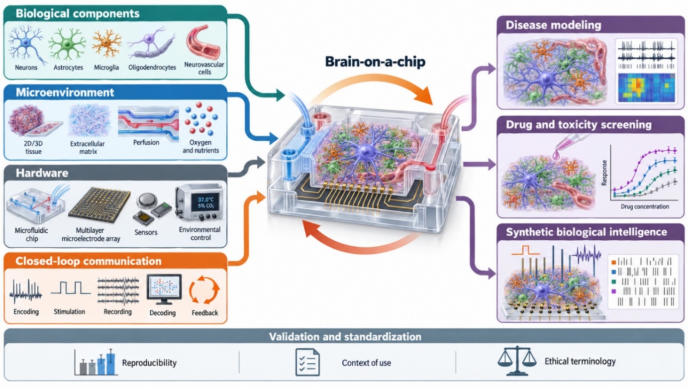
Brain-on-a-chip systems are constructed through coordinated design of four interconnected layers: biological components, including neurons, astrocytes, microglia, oligodendrocytes, and neurovascular cells; the cellular microenvironment, including two- or three-dimensional organization, extracellular matrix, perfusion, oxygen, and nutrients; hardware, including microfluidic devices, multilayer microelectrode arrays, sensors, and environmental controls; and closed-loop communication, in which biological activity is encoded, stimulated, recorded, decoded, and returned as feedback. Integration of these layers enables applications in disease modeling, drug and toxicity screening, and synthetic biological intelligence. The reliability and interpretation of each application depend on reproducibility, a clearly defined context of use, standardized validation criteria, and terminology that avoids unsupported anthropomorphic claims.
Biological Components of BoC
2.1. Selecting biological complexity by function
The mechanism and endpoint under study determine the minimum biological composition. Network electrophysiology may require reproducible excitatory and inhibitory neuronal populations. Myelination requires oligodendrocytes in direct contact with axons. Neuroinflammation requires microglia with a validated homeostatic baseline and stimulus-responsive phenotype. A BBB model requires endothelial cells, pericytes, astrocytic support, a basement-membrane-like interface, and directional vascular-tissue organization. Adding every available lineage is not automatically advantageous: excess complexity can reduce reproducibility, obscure causal interpretation, and introduce incompatible media requirements.
Cell source determines the scope of inference. Human iPSCs and embryonic stem cells provide renewable, genetically manipulable sources for regional and lineage differentiation. Patient-specific iPSCs enable genotype-associated phenotyping, particularly when paired with gene-corrected or engineered isogenic controls. Primary animal cells often show robust short-term function and naturally occurring cellular mixtures, but introduce species differences and finite supply. Immortalized lines improve convenience but may lose specialized functions. These sources are complementary, not interchangeable.
2.2. Neurons
Neurons provide the electrically excitable core of most BoC models. They generate action potentials, form synapses, integrate stimulation, and support readouts of excitability, connectivity, plasticity, neurotoxicity, and network adaptation. Selection of neuronal identity depends on the experimental question: cortical glutamatergic neurons are useful for network electrophysiology; GABAergic neurons are needed to examine excitation-inhibition balance; dopaminergic neurons are relevant to selected Parkinson’s disease mechanisms; and motor, sensory, or autonomic neurons are required for corresponding circuits.
Pluripotent-stem-cell differentiation generally follows developmental patterning or forced transcription-factor programming. Dual-SMAD inhibition suppresses non-neural fates and produces neural progenitors that can subsequently be patterned with morphogens [6,7]. This route offers regional flexibility and partially follows developmental signaling logic, but it is slower and vulnerable to line- and batch-dependent variation. Forced expression of transcription factors such as NGN2 produces relatively homogeneous excitatory neurons rapidly and reproducibly [8]. NGN2-derived neurons are therefore widely used in MEA studies, but rapid differentiation does not imply cortical completeness: excitatory-dominant cultures omit neuronal diversity, developmental trajectories, and often mature glial support.
Primary neurons dissociated from rodent brain provide an alternative for stable network studies [9,10]. They may retain naturally derived neuronal-glial mixtures, but ratios depend on source region, developmental age, dissociation, and culture conditions. Primary cultures cannot establish human-specific responses, and dissociation disrupts native architecture. For every source, spontaneous spikes alone are insufficient evidence of maturity. Rigorous characterization includes reporting subtype composition, excitatory-to-inhibitory ratio, differentiation efficiency, culture age, synaptic markers, pharmacological responsiveness, firing and bursting features, and predetermined functional acceptance criteria.
2.3. Astrocytes
Astrocytes regulate extracellular ions and neurotransmitters, provide metabolic and trophic support, influence synapse formation and plasticity, and contribute to neurovascular homeostasis. Their presence can alter neuronal survival, network synchrony, drug response, and disease phenotype. Consequently, a neuron-only preparation constitutes a neuronal-network model rather than a complete brain-tissue model.
iPSCs-derived astrocytes can be generated through neural-progenitor, neurosphere, and long-term organoid routes. Reported protocols differ markedly in purity, maturation time, regional identity, and reactive state [11,12]. Primary rodent astrocytes can be highly enriched by immunopanning [13], but species and developmental-stage differences remain. Serum exposure, dissociation, stiff substrates, and inflammatory cues can drive reactive phenotypes; lineage-marker characterization therefore requires functional assays of glutamate uptake, potassium handling, calcium signaling, metabolic support, and stimulus-specific reactivity.
Recent evidence directly supports the importance of astrocyte-derived signals in functional maturation: astrocyte-conditioned medium increased activity and synchrony in human forebrain organoids measured by MEA, although the magnitude and mechanism remained context dependent [14]. This result reinforces a general design principle: glial support is not a decorative addition but an experimental variable that can change the electrophysiological endpoint.
2.4. Microglia
Microglia are yolk-sac-derived resident immune cells that mediate surveillance, cytokine signaling, phagocytosis, synaptic remodeling, and responses to injury and protein aggregates. They are required for models of neuroinflammation and many neurodegenerative mechanisms. Because they arise from primitive myeloid rather than neuroectodermal progenitors, their separate derivation is necessary before integration into neural cultures [15].
Microglia are particularly sensitive to isolation, serum, substrate, cytokines, cell death, and medium composition. An apparently activated phenotype may reflect the model rather than the disease stimulus. Validation encompasses homeostatic and activation-state markers, ramified morphology, phagocytosis, cytokine profiles, and stimulus-specific responses. Microglia-containing organoids have demonstrated interactions with synaptic materials and changes in neuronal activity, but integration alone does not prove physiological maturation [16].
A 2026 study assembled separately differentiated iPSCs-derived cortical neurons, astrocytes, and microglia into a reproducible, postmitotic three-dimensional tissue that remained viable for months without a necrotic core and supported relatively homeostatic microglial phenotypes. The platform also reproduced selected Alzheimer’s-associated features after APP engineering or amyloid exposure [17]. This engineered-assembly strategy may offer greater compositional control than spontaneous organoid differentiation, although it sacrifices some developmental self-organization.
2.5. Oligodendrocytes
Oligodendrocytes form central nervous system myelin and provide metabolic support to axons. They are necessary for studies of conduction, white-matter injury, demyelination, and remyelination. Rapid protocols use transcription factors such as SOX10, OLIG2, and NKX6.2 to direct pluripotent-stem-cell-derived neural progenitors toward oligodendrocyte lineages [18,19]. Developmentally guided 3D cultures can also generate oligodendrocytes and support interactions with neurons [20]. These protocols generate oligodendrocytes from neural progenitors rather than hematopoietic stem cells.
The oligodendrocyte lineage marker O4 or other lineage-marker expression does not demonstrate functional myelination. Functional validation assesses axonal ensheathment, compact myelin, nodal organization, metabolic support, and changes in conduction velocity. Separately produced oligodendrocytes may survive poorly or fail to integrate after transfer, making assembly timing and direct axonal contact critical design variables.
2.6. Neurovascular cells
The BBB is generated by specialized brain microvascular endothelial cells interacting with pericytes, astrocytes, neurons, basement membrane, and vascular flow. A clear distinction separates a simple endothelial permeability assay from a neurovascular-unit model. Appropriate validation includes continuous junctional organization, transendothelial electrical resistance where applicable, size-selective permeability, transporter activity, directional polarity, and response to inflammatory or hypoxic perturbation.
Stem-cell-derived BBB models offer patient specificity but vary in endothelial identity and maturity. Primary cells can provide stronger barrier function but have limited availability; immortalized cells are convenient yet frequently form weaker barriers. Modular systems may permit separately matured tissues to be combined at an appropriate time. For example, a BBB-in-a-CUBE model integrated astrocytes, pericytes, and iPSCs-derived brain microvascular endothelial-like cells and assessed junctional markers, electrical resistance, passive permeability, and transporter-dependent drug behavior [21].
2.7. Multicellular assembly and acceptance criteria
Multicellular BoC systems can be generated by mixed differentiation, sequential addition, fusion of regional organoids, separately differentiated cell assembly, transwell coculture, or microfluidic compartmentalization. Direct contact is required for mechanisms such as myelination and many synaptic or phagocytic interactions; shared medium alone is sufficient only for soluble signaling questions. Because each lineage may require different maturation times and media, modular assembly can improve control but introduces handling and integration artifacts.
Before an experiment begins, each lineage requires prespecified identity and function criteria. The complete system additionally requires testing for cell survival, ratio stability, spatial distribution, baseline electrophysiology, inflammatory state, and responses to positive and negative controls. A model that does not meet baseline criteria is unsuitable for disease or computational interpretation.
Biological Microenvironment and Culture Configuration
3.1. Extracellular matrix and adhesion
Neurons and glia require an adhesive interface for survival, process extension, and network formation. Common coatings include poly-D-lysine, polyethylenimine, laminin, collagen, and Matrigel [22-25]. Positively charged polymers enhance attachment but do not reproduce brain extracellular matrix. Laminins and collagens provide receptor ligands, whereas Matrigel contains many bioactive components but is incompletely defined, animal-derived, and variable between lots. Coating density, stiffness, thickness, degradation, and spatial uniformity can alter neurite growth and electrode coupling.
3D scaffolds include natural or synthetic hydrogels, porous matrices, printed bioinks, and decellularized tissue-derived materials. A brain-extracellular-matrix-enhanced microfluidic system improved selected structural and functional features of brain organoids and used periodic flow to support survival and reduce variability [26]. These benefits are counterbalanced by batch variability, uncertain composition, and difficulty separating matrix effects from flow or geometry.
3.2. Media, oxygen, nutrients, and waste
Multicellular cultures require a medium that supports several lineages without selectively destabilizing another. Neuronal maturation supplements may not preserve endothelial barrier properties, and cytokines needed for microglial survival can alter neuronal or astrocytic states. A common medium is therefore a biological compromise. Compartment-specific perfusion, staged assembly, or controlled mixing can reduce this problem.
Small fluid volumes amplify evaporation, osmolarity drift, nutrient depletion, and accumulation of metabolites. 3D tissues additionally develop diffusion gradients and may form hypoxic or necrotic cores. Reliable operation requires monitoring or tight control of oxygen, glucose, lactate, pH, osmolarity, temperature, carbon dioxide, flow, and medium-exchange history. Physiological gradients can be intentional experimental variables; uncontrolled gradients are confounders.
3.3. Two-dimensional culture
Two-dimensional (2D) monolayers offer direct optical access, efficient exchange with medium, strong electrode coupling, established analysis methods, and relatively high throughput. They are well suited to causal electrophysiology, toxicity screening, and experiments requiring controlled neuronal composition. Their limitations include nonphysiological topology and stiffness, restricted three-dimensional connectivity, and incomplete cell-matrix organization. Human cells improve species relevance but do not remove immaturity, missing cell types, or absent systemic physiology.
3.4. Engineered 3D tissues and organoids
3D neural models include scaffold-based cultures, spheroids, patterned tissues, bioprinted constructs, self-organizing organoids, assembloids, and engineered assemblies. These forms are not interchangeable. Organoids exhibit self-organization and lineage or regional features; assembloids combine distinct organoid or cellular modules; engineered tissues prioritize compositional and geometric control.
3D organization can improve cell-cell and cell-matrix interactions, spatial patterning, and network topology, but complicates oxygen delivery, imaging, compound exposure, and electrical recording. It can also increase batch variability. A recent quality-control framework for cortical organoids assessed morphology, growth, cellular composition, cytoarchitecture, and cytotoxicity in a hierarchical workflow [27]. This type of standardized release testing is essential if organoid-based chips are to support reproducible screening.
3.5. Dynamic and time-evolving systems
The term “4D culture” is used inconsistently and does not denote a category inherently more advanced than 3D culture. A dynamic system is instead defined by its time-dependent variable: perfusion, changing biochemical gradients, programmed stimulation, matrix degradation, tissue morphing, maturation, or interaction with connected tissue modules [28]. A perfused transwell is not automatically a 4D culture. Complete characterization specifies the biological purpose, time scale, and measured consequence of each changing parameter.
Device Materials and Chip Design
4.1. Substrates, membranes, and compound compatibility
Common chip materials include glass, silicon, polystyrene, cyclic olefin polymers, polymethyl methacrylate, and polydimethylsiloxane (PDMS). PDMS offers optical transparency, gas permeability, and ease of prototyping, but can absorb hydrophobic compounds and release uncrosslinked oligomers. Its gas permeability and molecular sorption are bulk material properties, distinct from deliberately engineered pores. Material selection therefore involves tradeoffs among optical access, stiffness, bonding, sterilization, electrode fabrication, compound recovery, and manufacturing scalability.
Recent analysis of OoC adoption highlights material limitations, throughput, reproducibility, automation, and ease of use as factors influencing implementation beyond specialist engineering laboratories [29]. Microfabrication advances alone do not ensure adoption. Practical utility also depends on a defined intended use and a feasible quality-control workflow.
4.2. Microfluidic and perfusion architecture
Microfluidic architecture governs flow distribution, concentration gradients, sampling, compound residence time, shear stress, and waste removal. Neural compartments generally benefit from low shear, whereas vascular compartments may require physiologically relevant shear to support endothelial phenotype. System characterization includes channel dimensions, flow source, measured flow rate, recirculation configuration, reservoir volume, tubing materials, sampling losses, and compound recovery. Bubble traps and effective priming help prevent flow interruption, cellular damage, and electrical artifacts.
Perfusion enhances mass transport but can also alter cellular exposure through adsorption, dilution, or incomplete mixing. Computational fluid dynamics can inform device design, while experimental tracer measurements provide validation of residence times and concentration distributions. Reporting measured flow conditions supports reproducibility and interpretation more effectively than describing perfusion simply as “continuous circulation.”
Perfusion improves mass transport but can create unrecognized exposure differences through adsorption, dilution, or incomplete mixing. Computational fluid dynamics can guide initial design, but experimental tracer measurements are needed to validate residence time and concentration fields. Flow constitutes a measured input rather than an unquantified condition described simply as “continuous circulation.”
4.3. MEA physical architecture
MEAs contain electrodes distributed across a culture substrate for recording and, in some systems, stimulation. Their performance depends on electrode material, diameter, spacing, density, impedance, noise, reference configuration, amplifier characteristics, stimulation capability, and tissue coverage. Spatial resolution depends on these factors and electrode–tissue coupling, rather than channel count alone. Commercial and custom platforms range from arrays with tens of electrodes to high-density complementary metal-oxide-semiconductor (CMOS) arrays containing thousands of addressable sites [30].
Planar MEAs provide stable interfaces with neural monolayers but primarily record activity near the substrate. Three-dimensional interfaces, including penetrating shanks, mesh arrays, flexible wraps, and layered structures, extend access to deeper tissue regions. A multilayered MEA reported in 2026 incorporated stacked mesh electrode layers separated by compliant PDMS spacers to record activity at multiple depths within cerebral organoids [31]. This design expands spatial coverage while introducing challenges in assembly, electrode–tissue contact, and comparison of signals across layers.
Extracellular electrodes detect voltage changes generated by transmembrane ionic currents. These signals do not represent a bulk increase in extracellular ion concentration during an action potential. The reference electrode supplies a comparison potential for differential recording, while ground and stimulation-return electrodes serve distinct functions determined by the system architecture. Reporting electrode impedance, noise floor, reference configuration, and stimulation artifacts supports assessment of recording quality and interpretation of neural activity.
4.4. Housing and environmental control
Effective device housing supports sterility and stable temperature, carbon dioxide, pH, humidity, oxygenation, and osmolarity while limiting evaporation and maintaining optical and electrical access. Long-term operation depends on reliable seals, biocompatible tubing and adhesives, incubation-compatible connectors, and practical access for medium exchange and sampling. Ports that minimize contamination and pressure fluctuations further support culture stability.
Environmental monitoring is often less developed than electrophysiological recording. Integrated temperature, oxygen, pH, and metabolite sensors can help distinguish responses to experimental stimulation from changes associated with deteriorating culture conditions. Designs that allow cell or tissue retrieval also facilitate imaging, transcriptomic analysis, histology, and biochemical validation.
4.5. Assembly, quality control, and failure modes
Device qualification assesses dimensional accuracy, bonding integrity, resistance to leakage and pressure, sterility, susceptibility to bubble-related disruption, coating uniformity, flow distribution, electrode impedance, electrical noise, stimulation safety, evaporation, and compound recovery. After cell seeding, biological acceptance criteria provide an additional basis for determining experimental suitability. Potential failure modes include channel blockage, delamination, bubble formation, electrode corrosion, loss of cell adhesion, microbial contamination, reference-potential drift, shifts in cell-type proportions, tissue necrosis, and compound depletion through interactions with device materials.
Communication and Interface Systems
5.1. Information flow
A closed-loop BoC system comprises an ordered information pathway linking the experimental state to an encoder, stimulator, biological network, recording electrodes or sensors, amplifier and digitizer, preprocessing, feature extraction, decoder, software action, and feedback (Figure 1). Interpretation depends on explicit characterization of each transformation along this pathway. The biological network receives spatial and temporal patterns of electrical, optical, chemical, or mechanical stimulation defined by the encoding scheme, rather than abstract task variables such as “ball position.”
5.2. Input encoding and stimulation
Reproducible electrical stimulation depends on documentation of electrode location, pulse amplitude, phase width, interphase interval, frequency, waveform, train duration, charge balance, and return path. Charge-balanced biphasic pulses reduce net charge delivery, although safe stimulation limits depend on electrode material, surface area, and interface conditions. Stimulation artifacts can obscure immediate neural responses; blanking, artifact subtraction, hardware design, and predefined analysis windows offer complementary approaches to managing this interference.
Distinct encoding of environmental variables helps separate their contributions to network responses. Position, velocity, error, event identity, and performance feedback convey different information. Electrode location can encode spatial position, pulse rate or timing can represent continuous magnitudes, and stimulation patterns can distinguish categories. Establishing the discriminability of these inputs provides a basis for interpreting subsequent learning-related outcomes.
5.3. Signal acquisition and preprocessing
Documentation of signal acquisition includes sampling rate, analog and digital filters, input range, gain, noise levels, electrode impedance, synchronization, and associated metadata. Preprocessing can involve baseline correction, line-noise removal, stimulation-artifact handling, spike detection, optional spike sorting, and extraction of lower-frequency field activity. Prespecified or cross-validated detection criteria reduce the risk of inflating apparent effects through thresholds selected after examining outcomes.
Extracellular spikes reflect nearby transmembrane currents and may originate from one or multiple neurons. Changes in signal amplitude can arise from tissue movement, altered cell adhesion, electrode impedance, or cell death, complicating their interpretation as changes in neural information processing. Recordings from acellular chips, pharmacologically silenced cultures, and appropriate reference conditions help distinguish biological activity from device and stimulation artifacts.
5.4. Feature extraction and biological interpretation
Common electrophysiological measures include firing rate, interspike intervals, burst frequency and duration, synchrony, stimulus–response latency, evoked-response probability, estimated functional connectivity, and their changes over time. No single measure establishes learning. Evidence for learning depends on an explicit operational definition, appropriate baselines, within-culture controls, replication across independent cultures, and demonstration that performance gains depend on feedback rather than elapsed time, repeated exposure, or selective analysis windows.
Recorded voltage reflects electrophysiological activity; task actions arise through a decoder that maps predefined neural features to outputs. Reporting decoder performance with uncertainty estimates and comparisons against shuffled labels, replayed signals, and simple nonadaptive baselines helps distinguish neural adaptation from properties of the decoding procedure. Closed-loop latency also affects interpretation, as feedback delays can alter the relationship between neural activity, actions, and subsequent stimulation.
5.5. Feedback, reward, and anthropomorphic language
DishBrain demonstrated changes in task performance when cultured neurons were coupled to a simulated Pong environment through structured closed-loop feedback [32]. The study used predictable and unpredictable stimulation within an active-inference framework, but did not establish a universal neuronal preference for 100 Hz stimulation or a subjective experience of random stimulation as punishment. Although high-frequency stimulation can induce long-term potentiation in some preparations, its effects depend on cell type, developmental stage, stimulation intensity and pattern, and network state [33-35]. Terms such as “reward,” “punishment,” and “preference” therefore require explicit operational definitions, while claims of sentience require evidence beyond task performance.
More recent work reported feedback-dependent adaptation in mouse cortical organoids during a virtual pole-balancing task, extending closed-loop experiments beyond monolayer cultures [36]. Performance improved with adaptive training relative to random or null stimulation, but the improvement did not persist after a 45-min rest period. These results indicate task-specific adaptation with limited demonstrated retention; they do not establish consciousness, subjective experience, or general intelligence.
5.6. Software architecture, controls, and reporting
A reproducible software architecture preserves raw data, timestamps, stimulation events, analysis configurations, decoder versions, environmental metadata, and culture identifiers. Clearly distinguishing real-time processing from offline analysis supports reconstruction of experimental decisions. Machine-readable protocols, version-controlled code, documented random seeds, and access to representative raw data further support reproducibility.
Control conditions can include sham stimulation, acellular chips, viable cultures disconnected from feedback or operated in open loop, pharmacologically silenced cultures, replayed biological signals, and shuffled feedback. Matching total delivered charge across relevant stimulation conditions helps distinguish feedback effects from differences in stimulation exposure. Comparisons with artificial algorithms depend on comparable observations, available actions, training opportunities, feedback, and clearly defined computational budgets. Greater sample efficiency in a specific task alone does not establish general computational superiority over deep reinforcement learning methods.
Applications and Disease Models
6.1. Neurotoxicity and drug screening
BoC platforms combine repeated functional measurements with controlled exposure to detect changes that precede cell death. Relevant endpoints include network activity, calcium dynamics, barrier integrity, morphology, metabolism, inflammatory signaling, and viability. Their interpretation depends on a defined context of use, such as hazard identification, concentration–response analysis, mechanistic investigation, or prediction of clinical toxicity. Characterizing adsorption to device materials, protein binding, fluid volume, and exposure duration helps establish cellular exposure, which may differ substantially from the nominal concentration.
6.2. Blood-brain barrier transport
BBB chips can assess passive permeability, transporter-mediated flux, antibody and nanocarrier delivery, and inflammation-associated barrier disruption. Physiological interpretation depends on distinct vascular and tissue compartments, defined transport direction, and evidence of functional intercellular junctions and transport mechanisms. High transendothelial electrical resistance alone does not establish BBB fidelity, as a tight barrier can form without the characteristic transporter profile of brain endothelial cells. Surface-area-normalized permeability measurements and reference tracers of defined molecular sizes support comparisons across platforms.
6.3. Neurodegenerative disease
The validity of disease models depends on alignment between the initiating mechanism, vulnerable cell populations, spatial organization, and measured phenotypes. In Parkinson’s disease, dopaminergic neurons are particularly relevant to mechanisms underlying nigrostriatal degeneration, while astrocytes, microglia, pericytes, and endothelial cells enable investigation of specific glial and vascular contributions. A human substantia nigra brain chip incorporating dopaminergic neurons and neurovascular cells has been used to investigate α-synuclein-associated pathology and blood–brain barrier disruption [37]. Exposure to exogenous α-synuclein fibrils models selected downstream pathological processes but does not reproduce spontaneous, age-dependent disease initiation.
For Alzheimer’s disease, model interpretation depends on the source of amyloid and tau, genetic background, cellular aging state, neuroimmune composition, and vascular function. Patient-derived cells paired with isogenic controls can strengthen causal inference, although reprogramming to induced pluripotent stem cells can erase age-associated cellular features. Depending on the mechanism under investigation, approaches such as induced aging, prolonged maturation, defined inflammatory conditions, or direct conversion may help reproduce relevant aspects of the disease.
6.4. Validation framework
Validation of BoC platforms encompasses construct, phenotypic, and predictive validity. Construct validity concerns whether the initiating mechanism, cellular composition, spatial organization, and exposure conditions adequately represent the biological processes under investigation. Phenotypic validity assesses correspondence with relevant human molecular, structural, electrophysiological, inflammatory, and barrier abnormalities. Predictive validity evaluates whether responses to interventions agree with established human outcomes or, where human evidence is unavailable, well-supported preclinical findings, including the direction and magnitude of effects.
Translation also depends on operational performance, including within-run repeatability, reproducibility across days and operators, consistency between lots, transferability across laboratories, compatibility with automation, and predefined acceptance criteria. A clearly defined context of use links these validation measures to the intended regulatory or pharmaceutical decision, providing a more informative basis for evaluation than a general claim of human relevance.
Synthetic biological intelligence and multidimensional encoding
7.1. Scope of synthetic biological intelligence
SBI investigates adaptive information processing in living neural systems coupled to external environments. Although SBI shares components with disease-oriented BoC platforms, including neurons, microelectrode arrays, perfusion, stimulation, and software, its primary endpoint is feedback-dependent task performance rather than disease modeling or prediction of treatment responses.
Cultured neural networks can exhibit plasticity, adaptation, source separation, and task-related changes [32,38]. These findings support investigation of learning-like dynamics and the potential for energy-efficient biological computation, but do not establish that neural cultures can broadly replace artificial intelligence, digital computers, or semiconductor hardware. Comparisons with semiconductor technologies also require consideration of the physical, engineering, and economic constraints on scaling, including quantum tunneling, leakage currents, heat dissipation, interconnect delays, device variability, and manufacturing costs. Attributing these constraints broadly to the Heisenberg uncertainty principle oversimplifies their underlying mechanisms.
7.2. A testable multidimensional encoding framework
A multidimensional game-world paradigm offers a testable framework for examining whether living neuronal networks can integrate independently encoded environmental variables and whether adaptation to contingent feedback generalizes as additional variables are introduced. These capabilities remain research hypotheses rather than established properties of neural cultures.
NGN2-derived excitatory neurons offer rapid generation and reproducibility, while defined populations of inhibitory neurons and astrocytes may support investigation of a broader range of network dynamics. Network composition and electrophysiological characterization provide a basis for interpreting subsequent task performance. A 64-channel array is one possible starting configuration, with the allocation of recording and stimulation channels determined by platform capabilities. Channels designated by the manufacturer for reference or system functions remain unavailable for reassignment.
7.3. Encoding Strategy
Four nonoverlapping stimulation regions could encode distinct environmental variables, with separate recording regions providing candidate control outputs. Initial characterization would assess the discriminability and stability of responses evoked by each variable independently. Spatial position could be represented by electrode location, whereas continuous variables, such as distance or velocity, could be encoded through pulse rate, timing, or stimulation pattern. Separating performance feedback from sensory encoding would facilitate interpretation of network responses.
Progressively narrowing a random-frequency range around 100 Hz would alter frequency variability and temporal predictability and, depending on implementation, could also change mean frequency and delivered charge. These changes would confound attribution of performance gains to feedback contingency. A more controlled approach would compare prespecified conditions with matched total delivered charge across active stimulation groups. These conditions could include predictable feedback contingent on correct actions, identical feedback replayed noncontingently, contingent feedback with shuffled spatial assignments, and open-loop sensory stimulation without performance feedback. Sham stimulation and pharmacological silencing would provide additional controls for stimulation effects and dependence on neuronal activity.
A stimulation pattern could be described as reinforcing only if it produces reproducible improvements in task performance that depend on feedback contingency relative to appropriate controls. The primary endpoint could be the change in task success relative to open-loop and noncontingent replay conditions. Secondary endpoints could include response latency, evoked-response reliability, burst structure, functional connectivity, and retention after an interval without feedback.
7.4. Multiple-variable learning
Sequential introduction of variables begins only after performance for the first variable exceeds a prespecified threshold. The critical test is not whether activity changes, but whether information from two variables improves action selection compared with either variable alone. Factorial task designs can separate integration from simple cue dominance. Cross-validation on held-out task states and blinded analysis reduce the risk of overinterpretation.
If performance deteriorates when a new variable is added, possible explanations include limited discriminability, stimulation interference, decoder instability, network fatigue, or catastrophic interference. Testing these alternatives is necessary before attributing the decline to limited multisensory capacity. More electrodes or a 3D culture may increase interface capacity, but neither guarantees improved information integration.
Research gaps and future directions
8.1. Maturation and aging
Stem-cell-derived neural models generally resemble fetal or early postnatal tissue more closely than the adult brain. Long-term culture, astrocytic support, patterned activity, metabolic conditioning, and 3D organization can enhance selected aspects of maturation, but these approaches do not consistently establish an adult phenotype. Modeling age-associated diseases therefore depends on strategies that reproduce relevant features of aging, with their validity assessed against human reference datasets.
8.2. Reproducibility and standardization
Reproducibility depends on reporting cell sources and lines, passage numbers, differentiation protocols, cell-type ratios, culture age, matrix lots, and medium composition, together with device materials, geometry, flow conditions, electrode configurations, acquisition settings, preprocessing methods, and exclusion criteria. For organoid studies, characterization of morphology, cellular composition, cytoarchitecture, viability, and functional performance provides a basis for assessing suitability before experimental use. Reference compounds and shared datasets can further support comparisons across laboratories.
8.3. Interface completeness
Planar microelectrode arrays primarily record activity near the tissue–electrode interface, limiting coverage of deeper regions within 3D neural tissues. Penetrating and layered interfaces can extend recording coverage but may cause tissue damage or impose mechanical constraints. Integrating electrical recording with optical, chemical, metabolic, and environmental sensing could provide a more comprehensive assessment of tissue activity and culture conditions. The benefits of increased channel counts depend on effective electrode–tissue coupling, adequate signal-to-noise ratios, suitable data-processing methods, and broader spatial coverage.
8.4. Translation and context of use
The translational value of a BoC platform depends on a clearly defined intended use and evidence relevant to the decision it supports. For drug screening, key considerations include throughput, reproducibility, compound recovery, positive controls, and predictive performance. Disease models are evaluated through their mechanistic and phenotypic validity, whereas SBI platforms are assessed through defined tasks, activity-dependent feedback, appropriate baselines, and reproducible task performance. Claims tied to measured endpoints support more reliable interpretation than broad assertions of platform capability.
8.5. Ethics and terminology
Increasing complexity in closed-loop neural cultures and organoids raises ethical considerations concerning tissue provenance, donor consent, genetic manipulation, culture duration, stimulation protocols, and data governance. Current evidence does not establish consciousness in these systems, although uncertainty about their emerging capabilities supports the development of monitoring frameworks and explicit criteria for additional ethical review. Such frameworks can guide oversight as capabilities evolve. Anthropomorphic descriptions risk overstating these capabilities when they lack clear operational definitions and supporting evidence.
Conclusion
Brain-on-a-chip research is moving from isolated neural cultures toward multicellular, perfused, sensor-integrated, and closed-loop systems. The central challenge is not to reproduce every component of the human brain, but to construct the simplest model that faithfully represents the mechanism and endpoint of interest. Biological source and maturity, extracellular conditions, device materials, mass transport, electrode architecture, signal processing, and software feedback jointly determine validity.
Recent advances in microglia-containing tissues, organoid quality control, astrocyte-supported maturation, multilayer MEAs, modular BBB systems, and closed-loop organoid learning expand what can be measured and manipulated. They also expose new risks of confounding and overinterpretation. Progress will therefore depend on fit-for-purpose co-design, standardized acceptance criteria, transparent reporting, appropriate controls, and restraint in claims about human prediction or biological intelligence.
Abbreviations
2D: Two-dimensional
3D: Three-dimensional
4D: Four-dimensional
APP: Amyloid precursor protein
BBB: Blood–brain barrier
BoC: Brain-on-a-chip
GABA: Gamma-aminobutyric acid
iPSCs: Induced pluripotent stem cells
LTP: Long-term potentiation
MEA: Microelectrode array
NGN2:Neurogenin 2
NKX6.2: NK6 homeobox 2
OLIG2: Oligodendrocyte transcription factor 2
OoC: Organ-on-a-chip
PDMS: Polydimethylsiloxane
SBI: Synthetic biological intelligence
SMAD: SMAD family signaling protein
SOX10: SRY-box transcription factor 10
Declarations
Author Contributions
Conceptualization, C.P.C. and F.F.; formal analysis, C.P.C.; investigation, C.P.C.; writing original draft preparation, C.P.C.; writing review and editing, C.P.C.; visualization, C.P.C.; supervision, F.F. All authors have read and agreed to the published version of the manuscript.
Funding
This research received no external funding.
Institutional Review Board Statement
Not applicable.
Informed Consent Statement
Not applicable.
Data Availability Statement
Not applicable.
Conflicts of Interest
The authors declare no conflict of interest.
References
- Wheeler, B. C. Building a brain on a chip. Annu. Int. Conf. IEEE Eng. Med. Biol. Soc. IEEE Eng. Med. Biol. Soc. Annu. Int. Conf. 2008, 1604–1606 (2008). Google Scholar ↗
- Bang, S., Jeong, S., Choi, N. & Kim, H. N. Brain-on-a-chip: A history of development and future perspective. Biomicrofluidics 13, 051301 (2019). Google Scholar ↗
- Servais, B. et al. Engineering brain-on-a-chip platforms. Nat. Rev. Bioeng. 2, 691–709 (2024). Google Scholar ↗
- Smirnova, L. et al. Organoid intelligence (OI): the new frontier in biocomputing and intelligence-in-a-dish. Front. Sci. 1, (2023). Google Scholar ↗
- Pașca, S. P. et al. A framework for neural organoids, assembloids and transplantation studies. Nature 639, 315–320 (2025). Google Scholar ↗
- Chambers, S. M. et al. Highly efficient neural conversion of human ES and iPS cells by dual inhibition of SMAD signaling. Nat. Biotechnol. 27, 275–280 (2009). Google Scholar ↗
- Puspita, L., Deline, M. & Shim, J. Dual SMAD inhibition as a versatile platform in human pluripotent stem cell–based regenerative medicine and disease modeling. Mol. Cells 48, 100284 (2025). Google Scholar ↗
- Hulme, A. J., Maksour, S., Glover, M. S.-C., Miellet, S. & Dottori, M. Making neurons, made easy: The use of Neurogenin-2 in neuronal differentiation. Stem Cell Rep. 17, 14–34 (2022). Google Scholar ↗
- Yaron, A. et al. Dissociated neuronal cultures as model systems for self-organized prediction. Front. Neural Circuits 19, (2025). Google Scholar ↗
- Cano-Jaimez, M. et al. Preparation of dissociated mouse primary neuronal cultures from long-term cryopreserved brain tissue. J. Neurosci. Methods 330, 108452 (2020). Google Scholar ↗
- Zhou, S. et al. Neurosphere Based Differentiation of Human iPSC Improves Astrocyte Differentiation. Stem Cells Int. 2016, 4937689 (2016). Google Scholar ↗
- Sloan, S. A. et al. Human Astrocyte Maturation Captured in 3D Cerebral Cortical Spheroids Derived from Pluripotent Stem Cells. Neuron 95, 779-790.e6 (2017). Google Scholar ↗
- Foo, L. C. et al. Development of a Method for the Purification and Culture of Rodent Astrocytes. Neuron 71, 799–811 (2011). Google Scholar ↗
- Zheng, H. et al. Astrocyte-secreted cues promote neural maturation and augment activity in human forebrain organoids. Nat. Commun. 16, 2845 (2025). Google Scholar ↗
- Abud, E. M. et al. iPSC-Derived Human Microglia-like Cells to Study Neurological Diseases. Neuron 94, 278-293.e9 (2017). Google Scholar ↗
- Zhang, W. et al. Microglia-containing human brain organoids for the study of brain development and pathology. Mol. Psychiatry 28, 96–107 (2023). Google Scholar ↗
- Klimmt, J. et al. A reproducible three-dimensional model of human brain tissue to investigate physiological and disease-associated microglia phenotypes. Nat. Neurosci. 29, 2324–2340 (2026). DOI ↗ Google Scholar ↗
- Ehrlich, M. et al. Rapid and efficient generation of oligodendrocytes from human induced pluripotent stem cells using transcription factors. Proc. Natl. Acad. Sci. 114, E2243–E2252 (2017). Google Scholar ↗
- García-León, J. A. et al. SOX10 Single Transcription Factor-Based Fast and Efficient Generation of Oligodendrocytes from Human Pluripotent Stem Cells. Stem Cell Rep. 10, 655–672 (2018). Google Scholar ↗
- Marton, R. M. et al. Differentiation and maturation of oligodendrocytes in human three-dimensional neural cultures. Nat. Neurosci. 22, 484–491 (2019). Google Scholar ↗
- Koh, I. & Hagiwara, M. Modular tissue-in-a-CUBE platform to model blood-brain barrier (BBB) and brain interaction. Commun. Biol. 7, 177 (2024). Google Scholar ↗
- Hyvärinen, T. et al. Functional characterization of human pluripotent stem cell-derived cortical networks differentiated on laminin-521 substrate: comparison to rat cortical cultures. Sci. Rep. 9, 17125 (2019). Google Scholar ↗
- Passaniti, A., Kleinman, H. K. & Martin, G. R. Matrigel: history/background, uses, and future applications. J. Cell Commun. Signal. 16, 621–626 (2022). Google Scholar ↗
- Stil, A. et al. A simple method for poly-D-lysine coating to enhance adhesion and maturation of primary cortical neuron cultures in vitro. Front. Cell. Neurosci. 17, 1212097 (2023). Google Scholar ↗
- Young, E. W. K. & Beebe, D. J. Fundamentals of microfluidic cell culture in controlled microenvironments. Chem. Soc. Rev. 39, 1036–1048 (2010). Google Scholar ↗
- Cho, A.-N. et al. Microfluidic device with brain extracellular matrix promotes structural and functional maturation of human brain organoids. Nat. Commun. 12, 4730 (2021). Google Scholar ↗
- Castiglione, H. et al. Towards a quality control framework for cerebral cortical organoids. Sci. Rep. 15, 29431 (2025). Google Scholar ↗
- Miao, S. et al. 4D Self-Morphing Culture Substrate for Modulating Cell Differentiation. Adv. Sci. 7, 1902403 (2020). Google Scholar ↗
- Alver, C. G., Drabbe, E., Ishahak, M. & Agarwal, A. Roadblocks confronting widespread dissemination and deployment of Organs on Chips. Nat. Commun. 15, 5118 (2024). Google Scholar ↗
- Müller, J. et al. High-resolution CMOS MEA platform to study neurons at subcellular, cellular, and network levels. Lab. Chip 15, 2767–2780 (2015). Google Scholar ↗
- Kim, N. et al. Multilayered microelectrode array for monitoring electrophysiological signals of 3D neural networks in cerebral organoids. Microsyst. Nanoeng. 12, 201 (2026). DOI ↗ Google Scholar ↗
- Kagan, B. J. et al. In vitro neurons learn and exhibit sentience when embodied in a simulated game-world. Neuron 110, 3952-3969.e8 (2022). Google Scholar ↗
- Wang, H. & Wagner, J. J. Priming-Induced Shift in Synaptic Plasticity in the Rat Hippocampus. J. Neurophysiol. 82, 2024–2028 (1999). Google Scholar ↗
- Brown, G. P. et al. Long-Term Potentiation Induced by θ Frequency Stimulation Is Regulated by a Protein Phosphatase-1-Operated Gate. J. Neurosci. 20, 7880–7887 (2000). Google Scholar ↗
- Clapp, W. C., Hamm, J. P., Kirk, I. J. & Teyler, T. J. Translating LTP from animals to humans: A novel method for non-invasive assessment of cortical plasticity. Biol. Psychiatry 71, 496–502 (2012). Google Scholar ↗
- Robbins, A. et al. Goal-directed learning in cortical organoids. Cell Rep. 45, 116984 (2026). DOI ↗ Google Scholar ↗
- Pediaditakis, I. et al. Modeling alpha-synuclein pathology in a human brain-chip to assess blood-brain barrier disruption. Nat. Commun. 12, 5907 (2021). Google Scholar ↗
- Isomura, T., Kotani, K. & Jimbo, Y. Cultured Cortical Neurons Can Perform Blind Source Separation According to the Free-Energy Principle. PLOS Comput. Biol. 11, e1004643 (2015). Google Scholar ↗