
Abstract
Non-pigmenting fixed drug eruption (FDE) is a rare clinical variant characterized by the absence of residual hyperpigmentation, often leading to diagnostic confusion with other vesicobullous disorders. It differs from classical FDE in its atypical resolution pattern. Certain drugs, including fluoroquinolones such as ofloxacin, may be implicated in both non-pigmenting and pigmenting forms. Recognition of this spectrum, along with careful drug evaluation, is essential for accurate diagnosis, appropriate management, and prevention of recurrence. We report a case of fixed drug eruption induced by oral ofloxacin (200 mg), initially presenting as a non-pigmenting variant of FDE and subsequently evolving into the classical pigmenting variant of FDE upon re-exposure, highlighting its clinical course and importance in differential diagnosis; ofloxacin should be considered a potential causative agent for both non-pigmenting and pigmenting FDE.
Keywords
Ofloxacin Non-pigmenting fixed drug eruption Pigmenting fixed drug eruption oral drug rechallenge test.
Introduction
Fixed drug eruption (FDE) is a well-recognized cutaneous adverse drug reaction characterized by the recurrence of sharply demarcated lesions at the same sites upon re-exposure to the offending agent, typically resolving with residual hyperpigmentation. Non-pigmenting fixed drug eruption represents a rare clinical variant in which lesions heal without post-inflammatory hyperpigmentation, often posing diagnostic challenges by mimicking other vesicobullous dermatoses. This entity is likely underrecognized and underreported, as the absence of residual hyperpigmentation may limit patient awareness and delay clinical presentation. The pathogenesis involves a localized delayed-type hypersensitivity reaction mediated by resident memory T cells. In classical FDE, melanocyte activation and pigment incontinence lead to persistent hyperpigmentation, whereas in the non-pigmenting variant, altered melanocyte response or minimal basal keratinocyte damage may account for the lack of pigmentation.
Fluoroquinolones, including ofloxacin, are established causes of FDE; however, their association with the non-pigmenting variant remains infrequently reported.
Only a few cases of non-pigmenting fixed drug eruption (NPFDE) have been reported in the literature, and to the best of our knowledge, transition from non-pigmenting to pigmenting FDE upon re-exposure has not been previously described.
Case presentation
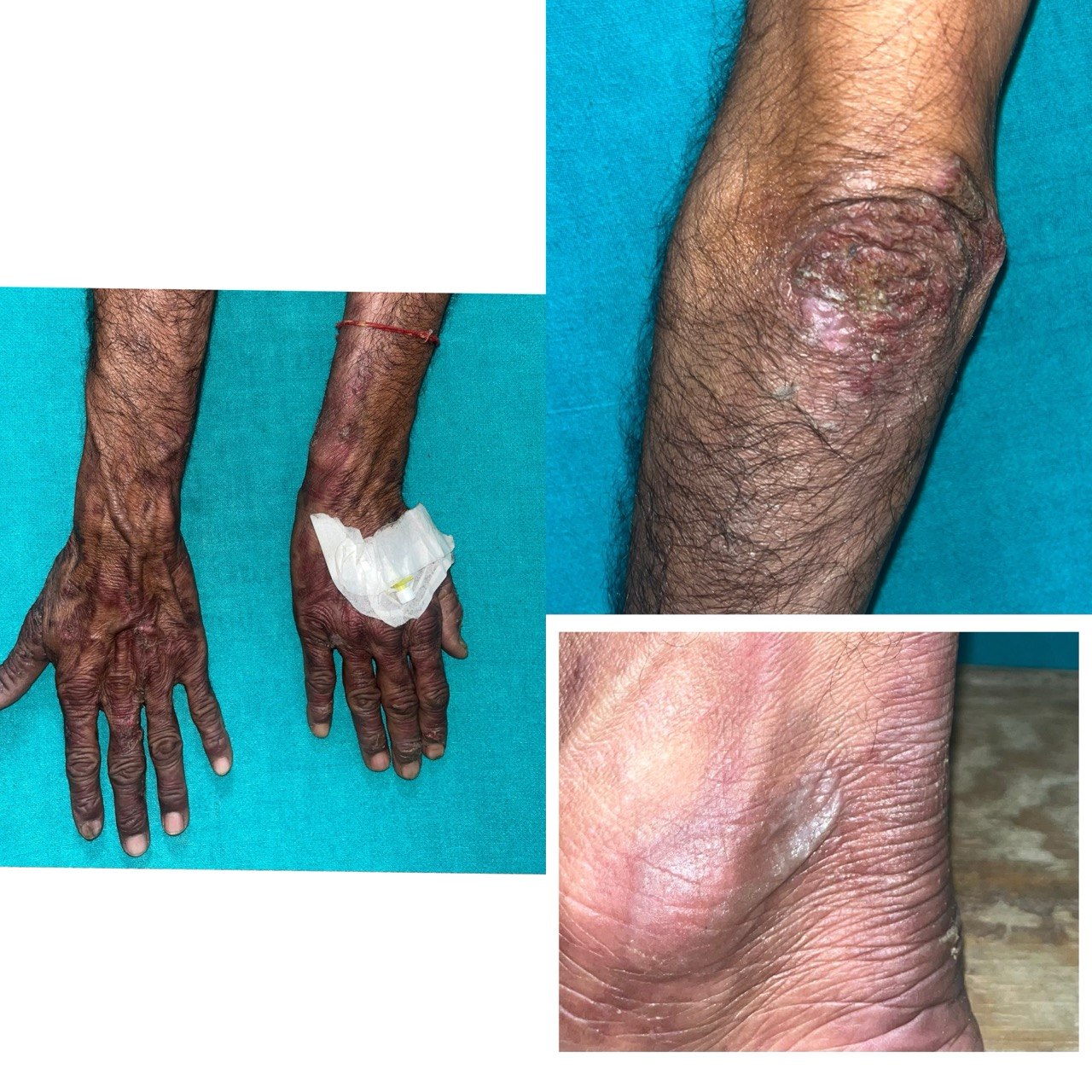
A 42-year-old male presented with a 10-day history of vesiculobullous lesions over the bilateral upper and lower limbs and buttocks. The lesions were preceded by erythematous, pruritic patches over the elbows, knees, and dorsum of the hands and feet, which progressed to blister formation within 2–3 days. The patient had taken oral ofloxacin (200 mg), prescribed by a physician for diarrhoea, 1–2 days prior to the onset of lesions.
On examination, well-defined erythematous plaques and bullae of varying sizes were noted over the extremities, predominantly distal to the elbows and knees, as well as over the buttocks. The lesions were tender, with blister formation at some sites. Routine laboratory investigations were within normal limits.
Histopathology showed basal cell vacuolization, lymphocytic exocytosis, subepidermal split, and interface dermatitis with perivascular infiltrate.
A diagnosis of non-pigmenting bullous fixed drug eruption was established based on clinical presentation, drug association, absence of residual hyperpigmentation, and histopathological findings.
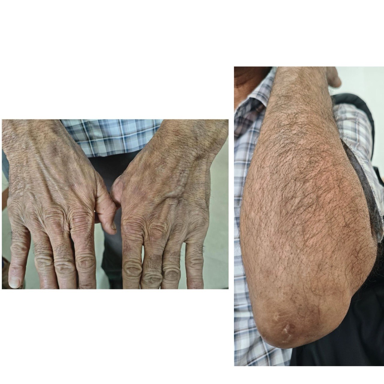
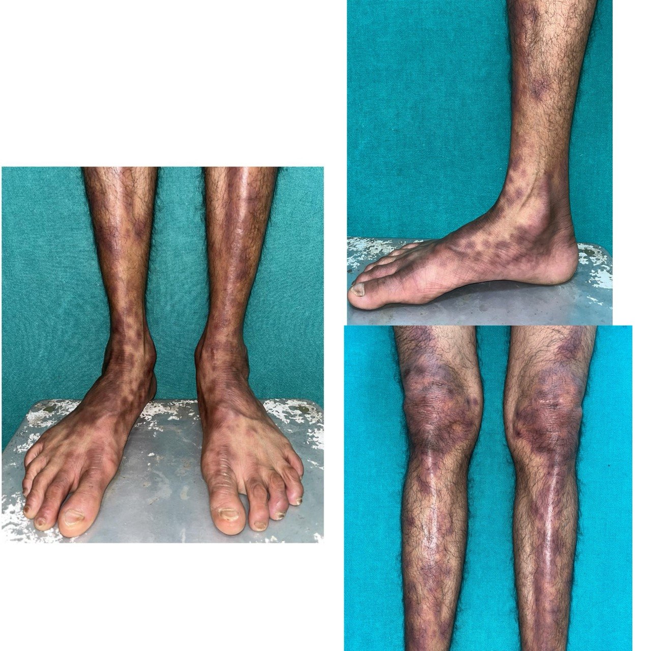
Withdrawal of the drug and short-course systemic corticosteroids led to complete healing within 10–15 days without pigmentation. The patient was advised strict avoidance of the offending drug. Six weeks after complete resolution of the lesions, an oral drug rechallange test with ofloxacin was performed. An initial quarter of the therapeutic dose was administered, and no reaction was observed even after a 2-week observation period. Subsequently, half of the therapeutic dose was given, within 12 hours, the patient developed localized pruritic, erythematous to violaceous patches and plaques at the sites of previously healed lesions. Additionally, new lesions appeared over the upper back, indicating both reactivation and involvement of new sites. No systemic symptoms were observed.
The lesions gradually subsided over the next few days, leaving behind residual hyperpigmentation, confirming the diagnosis of classical Fixed Drug Eruption. The recurrence of lesions at identical sites following oral drug rechallenge established the diagnosis of ofloxacin-induced FDE.
Results and Discussion
Non-pigmenting fixed drug eruption (FDE) is an uncommon variant characterized by the absence of residual hyperpigmentation following resolution. This contrasts with classical FDE, where post-inflammatory hyperpigmentation is a hallmark feature due to melanocyte activation and pigment incontinence. The underlying mechanism of non-pigmenting FDE remains unclear, but it may reflect minimal melanocyte stimulation, limited basal keratinocyte damage, or efficient clearance of inflammatory mediators, resulting in negligible melanin deposition in the dermis.
In the present case, the initial episode was consistent with non-pigmenting FDE; however, upon re-exposure to the offending drug, subsequent lesions healed with residual hyperpigmentation, indicating a transition from non-pigmenting FDE to the classical pigmenting variant of FDE. This transition suggests that repeated antigenic exposure may enhance cytotoxic T-cell–mediated damage to basal keratinocytes and melanocytes, leading to increased melanin release and dermal pigment incontinence. Additionally, cumulative inflammation may upregulate melanogenic cytokines (such as endothelin-1, α-MSH, and stem cell factor), promoting melanocyte activation and melanin synthesis in later episodes. The differential diagnoses included Stevens–Johnson syndrome and Erythema Multiforme. However, recurrence of lesions at identical sites after drug re-exposure, rapid onset following drug intake, well-demarcated vesiculobullous lesions, and absence of significant systemic symptoms favoured a diagnosis of fixed drug eruption. In contrast, Stevens–Johnson syndrome is associated with severe mucosal involvement and systemic symptoms, while erythema multiforme typically presents with classical target lesions and is commonly linked to herpes simplex infection.
Conclusion
This case highlights the rare occurrence of non-pigmenting fixed drug eruption and its potential evolution into the classical pigmenting variant upon re-exposure. It underscores the importance of careful drug history and supervised oral provocation testing in establishing the diagnosis. Additionally, it demonstrates that ofloxacin can act as a causative agent for both non-pigmenting and pigmenting FDE, reflecting variability in melanocyte response and disease expression.
Furthermore, variability in immune response and melanocyte activity may result in differing clinical expressions across episodes, with potential transition from non-pigmenting to pigmenting forms upon re-exposure.
Ofloxacin, a fluoroquinolone antibiotic, is a known cause of cutaneous adverse drug reactions. This case highlights that ofloxacin can act as a causative agent for both non-pigmenting and pigmenting variants of fixed drug eruption, reflecting the dynamic interplay between immune response and melanocyte activity in determining clinical presentation.
Declarations
Informed consent statement
Written informed consent was obtained from the next of kin for publication of this case report and accompanying images.
List of abbreviations
FDE: Fixed drug eruption
NPFDE: Non pigmenting – fixed drug eruption
Conflicts of Interest
There is no conflict of interest regarding the publication of this paper.
Funding Statement
No funding was received for conduction this study.
Authors' contributions
AJ - Design, concepts, manuscript preparation, manuscript editing
PC - Literature search, clinical studies, manuscript editing, manuscript review
NK - Data acquisition, data analysis, manuscript editing, manuscript review
DK - Clinical studies, data analysis, manuscript review
PR - Clinical studies, data analysis, manuscript review
All authors read and approved the final manuscript.
Acknowledgment
The authors would like to thank the Department of Dermatology for their clinical support and guidance in the diagnosis and management of this case. We also acknowledge the patient for providing consent for publication of the clinical details and images.
References
- Gupta LK, Beniwal R, Khare AK, Mittal A, Mehta S, Balai M. Non-pigmenting fixed drug eruption due to fluoroquinolones. Indian J Dermatol Venereol Leprol. 2017 Jan-Feb;83(1):108-112. doi: . PMID: 27647361. DOI ↗ Google Scholar ↗
- Das S, Das S, Chowdhury J, Bhanja DC. Non pigmenting mucosal fixed drug eruption due to tadalafil: A report of two cases. Indian Dermatol Online J. 2014 Apr;5(2):167-9. doi: . PMID: 24860753; PMCID: PMC4030346. DOI ↗ Google Scholar ↗
- Vidal C, Prieto A, Pérez-Carral C, Armisén M. Nonpigmenting fixed drug eruption due to pseudoephedrine. Ann Allergy Asthma Immunol. 1998 Apr;80(4):309-10. doi: . PMID: 9564979. DOI ↗ Google Scholar ↗
- Singhal RR, Sheth NK, Nair PA. Non-Pigmented Fixed Drug Eruption Caused by Ibuprofen. Indian Dermatol Online J. 2019 May-Jun;10(3):341-343. doi: . PMID: 31149592; PMCID: PMC6536071. DOI ↗ Google Scholar ↗
- Prabha N, Chhabra N. Non-pigmenting Fixed Drug Eruption: An Unusual Adverse Reaction to Aceclofenac. Curr Drug Saf. 2018;13(3):224-225. doi: . PMID: 30047334. DOI ↗ Google Scholar ↗