
Abstract
Introduction: Mast cells, comprising up to 29% of the inflammatory cell population, have been reported in periapical granulomas and periapical cysts. Through the release of mediators such as histamine, heparin, tryptase, chymase, and basic fibroblast growth factor, mast cells play a significant role in inflammation and tissue remodeling. Their identification is reliably achieved using metachromatic stains like Toluidine blue and Astra blue. The present study aimed to evaluate mast cell activity in periapical granulomas and periapical cysts using these staining techniques. Objective: 1. To stain the mast cells in Periapical granuloma and Periapical cyst using Toluidine blue and Astra blue stains 2. To compare the staining efficacy of Toluidine blue and Astra blue stains for mast cells. 3. To compare the number of mast cells between Periapical granuloma and Periapical cyst. Materials and Methods: This retrospective Cross-sectional study was conducted in the Department of Oral and Maxillofacial Pathology at Navodaya Dental College and Hospital, Raichur, India, from February 2024 to June 2025. A total of 80 cases (15-50 years) of Periapical lesions comprising 30 cases of Periapical Granuloma and 30 cases of Periapical Cyst. Chronic tonsilitis tissue served as positive controls (10 cases), Cleft lip and palatal tissue served as negative controls (10 cases). Ethical clearance for the study was obtained from the Institutional Ethics Committee. Tissue sections were stained with Toluidine blue and Astra blue for mast cell identification. Mast cells were identified by their metachromatic staining properties and were quantified under light microscopy in representative high-power fields (×400 magnification). and the data were statistically analyzed using ANOVA, followed by post hoc Dunnett’s t-test and unpaired t-test. Results: The mean mast cell counts in periapical granulomas were 52.6 ± 18.1 (Astra blue) and 49.5 ± 19.4 (Toluidine blue), whereas in periapical cysts they were 71.1 ± 31.7 and 69.9 ± 26.5, respectively. Comparison between periapical granulomas and periapical cysts using both stains revealed a statistically significant increase in mast cell count in cysts. Additionally, the mean mast cell count was higher in females than in males, irrespective of lesion type and staining method. Conclusion: On comparison, periapical cysts demonstrated a higher mast cell count than periapical granulomas. Astra blue was found to be more specific for mast cell identification compared to Toluidine blue. The findings of the present study re-emphasize the role of mast cells in the pathogenesis of chronic inflammatory periapical lesions. Clinically, these results suggest that therapeutic strategies targeting mast cell mediators may help modulate inflammation in such lesions.
Keywords
Mast Cells Immune Responses Toluidine blue Stain Astra blue stain Periapical Granulomas.
Introduction
Mast cells are known to play an important role in inflammatory and immune responses associated with periapical lesions. They release several mediators such as histamine, heparin, cytokines, and proteolytic enzymes that may contribute to tissue degradation, angiogenesis, and modulation of the inflammatory response. These mediators may influence the progression and expansion of periapical lesions, suggesting that mast cells may have a significant role in the pathogenesis of periapical granulomas and cysts [1]. Clinically, differentiation between periapical granuloma and periapical cyst is important because their biological behavior and treatment approaches may differ [2,3].
While many periapical granulomas respond well to conventional endodontic therapy, some periapical cysts, particularly true cysts, may persist and occasionally require surgical intervention. Therefore, understanding the cellular components involved in these lesions may help in explaining their pathogenesis and behavior. Accurate identification of mast cells is essential for evaluating their role in periapical lesions. Different histochemical stains demonstrate mast cells with varying sensitivity depending on the maturity and sulfation of granules [4]. Therefore, comparing staining techniques such as Toluidine Blue and Astra Blue is important for determining the most reliable method for mast cell detection and quantification in tissue sections [5].
Toluidine blue and other basic aniline dyes are used in metachromatic staining to demonstrate mast cells. Both the weakly and heavily sulfated types of mast cell granules can be stained with Astra blue, which is another very specific dye for demonstrating mast cells. Toluidine blue, on the other hand, exclusively stains mature mast cells [6]. Few studies have been conducted in the literature to quantify mast cells and assess their potential involvement in periapical granuloma and periapical cyst [5,7]. The findings from such studies may contribute to a better understanding of the inflammatory mechanisms involved in periapical pathology and may provide insights into the biological behavior of these lesions, potentially aiding in improved diagnostic interpretation and therapeutic planning by staining the mast cells.
Aims & Objectives
-
To stain the mast cells in Periapical granuloma and Periapical cyst using Toluidine blue and Astra blue stains
-
To compare the staining efficacy of Toluidine blue and Astra blue stains for mast cells.
-
To compare the number of mast cells between Periapical granuloma and Periapical cyst
Materials and Methods
This retrospective Cross-sectional study was conducted in the Department of Oral and Maxillofacial Pathology at Navodaya Dental College and Hospital, Raichur, India, from February 2024 to June 2025. Ethical clearance for the study was obtained from the Institutional Ethics Committee with IEC no: IEC/NDC/RCR/2024-2025/SS0012.
Inclusion and Exclusion Criteria
The study included histopathologically diagnosed cases of periapical granuloma and periapical cyst retrieved from the departmental of Oral and Maxillofacial Pathology, Navodaya Dental College and Hospital, Raichur. archives. Cases with well-preserved paraffin-embedded tissue blocks and confirmed diagnoses were included in the study. Cases with inadequate tissue samples, other periodontal lesions involving the tooth, and other odontogenic or non-odontogenic lesions not associated with pulpal origin were excluded.
Sample Size Estimation
The sample size was determined based on the availability of archived paraffin-embedded tissue blocks with confirmed histopathological diagnosis during the study period. The study included a total of 80 cases (15-50 years) of Periapical lesions comprising 30 cases of Periapical Granuloma and 30 cases of Periapical Cyst. Chronic tonsilitis tissue served as positive controls (10 cases), Cleft lip and palatal tissue served as negative controls (10 cases). Chronic tonsillitis tissue sections were used as positive controls because they represent chronic inflammatory lesions characterized by abundant inflammatory infiltrate, including mast cells, thereby validating the staining procedure for mast cell identification. Tissue samples from cleft lip and palate were used as negative controls, as these tissues generally demonstrate minimal inflammatory infiltrate and relatively fewer mast cells in the connective tissue [8,9]. These control tissues were selected from the departmental archives to ensure appropriate validation of the staining technique.
Methodology and Parameters Studied
Archived paraffin-embedded tissue blocks were retrieved from the departmental archives. Tissue sections of 3-4 µm thickness were obtained from the selected paraffin-embedded tissue blocks and sections were made using a microtome and mounted on glass slides for staining. One section from each block was stained with Hematoxylin and Eosin (H&E) to reconfirm the histopathological diagnosis. Additional sections were stained with Toluidine Blue and Astra Blue stains for mast cell identification. Mast cells were identified by their metachromatic staining properties with Toluidine blue and Astra blue stains and were quantified under light microscopy in representative high-power fields (×400 magnification).
Staining Procedure
The procedure followed is as per the instructions mentioned by Sharma R et al. (2010) [10]. The staining procedure involves the following steps in the study:
-
Toluidine blue staining
-
Sections were covered with toluidine blue solution (0.2 gm of Toluidine blue powder was dissolved in 100 ml of distilled water at pH 4.0) for 1 min followed by washing with tap water.
-
Differentiation in 100% ethyl alcohol was performed for 30 seconds.
-
Astra blue staining
-
Sections were covered with astra blue solution (1gm of Astra blue powder was dissolved in 100 ml of 0.7 N HCl at pH 0.3) for 45minutes, followed by differentiation in 0.7N HCl for 1 min.
-
After washing with water, counterstaining was done with 0.5% safranin for 2 seconds.
-
The sections were dehydrated increasing grades of alcohol for 2s per grade, cleared in xylene and later mounted using DPX.
Examination of Slides
Toluidine blue staining
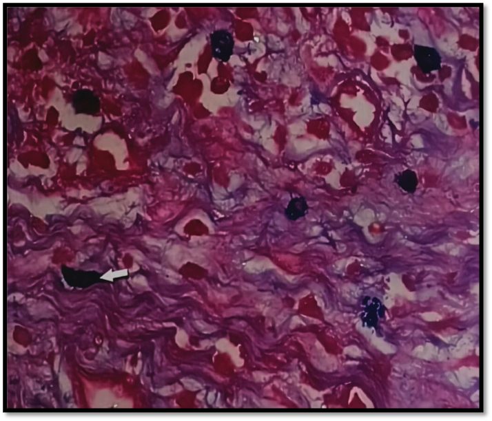
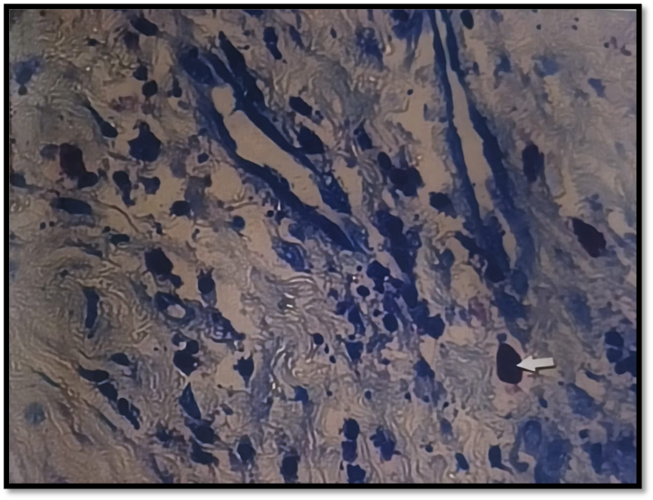
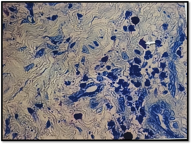
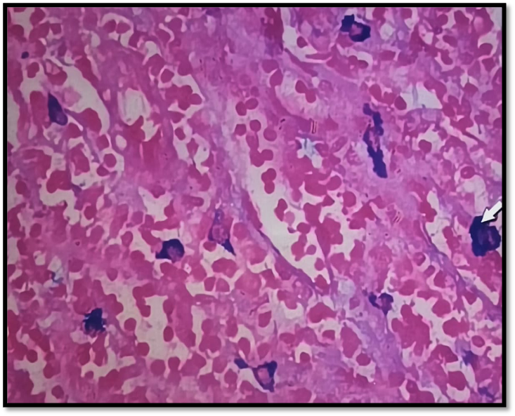
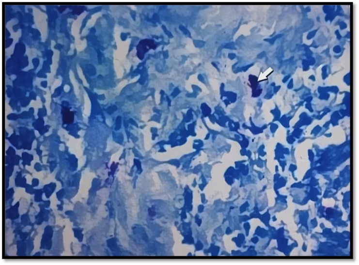
Mast cells appeared as oval or angular cells with purple granules and blue nuclei located mainly in the peripheral areas. Background tissue was stained in shades of blue [9,10].
Astra blue staining
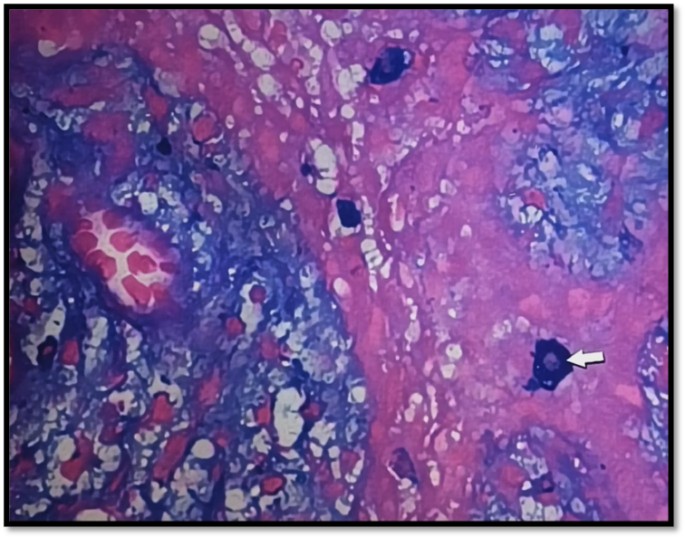
Mast cells appeared as oval or angular cells with blue granules and red nuclei located mainly in peripheral areas. Background tissue was stained in shades of red and blue [9,10].
Each section was evaluated for the number of mast cells and localization of mast cells in Periapical Granuloma and Periapical Cyst.
Counting procedure for number of mast cells
The Olympus BX53 Pentahead research microscope was used to analyse each slide at 40x magnification. Ten non-overlapping high-power fields (HPF) were selected from each stained section using a systematic method, when the slide is moved from right to left. Ten fields were photographed and saved to a computer, where mast cells were counted using a manual tag and an image analysis program (Image Pro-express). To correspond with the images' magnification, an icon called spatial calibration was used. Using the computer mouse, cells were counted by clicking on the corresponding tags. For each slide, the total number of cells was tallied over all ten photos, and care was taken to avoid field overlap. The outcomes were collated and subjected to statistical analysis.
Localization of Mast Cells
Localization of mast cells was also assessed. In Periapical Granuloma, presence of mast cells in center of the lesion was graded as “1”, periphery of lesion as “2” and near the vascularity as “3”. In Periapical Cyst, presence of mast cells in epithelium was graded as “1”, sub-epithelium as “2” and deeper connective tissue as “3” [9].
Two more observers carried out all these observations to eliminate inter-observer bias. The results were then tabulated and subjected to statistical analysis.
Statistical Analysis
The data was expressed in mean and SD. Comparison between the two study groups was done using Unpaired‘t’ test. Comparison between the study group and control group was done using ANOVA test for parametric distribution followed by post hoc Dunnett’s t-test. A two tailed P value > 0.05 is considered as “not significant” and P≤ 0.05 is considered as “significant”.
Results
Comparison of Periapical Granuloma and Periapical Cyst with Age & Gender
The prevalence of periapical granuloma and periapical cyst was compared with age using the Chi-square test. 13 of the 30 periapical granulomas were in patients under the age of 30, and 17 were in patients over 30. Of the 30 periapical cysts, 15 were found in individuals under 30 and 15 in those over 30. According to age, Table 1 indicates that the difference was not statistically significant (p-0.79).
Periapical granuloma and periapical cyst occurrences were compared by gender using the Chi-square test. Males accounted for 18 of the 30 cases of periapical granulomas, whereas females accounted for 12. Males accounted for 22 of the 30 cases of periapical cysts, while females accounted for 8. According to gender, Table 1, indicates there was no statistically significant difference (p-0.41).
| Age | Periapical Granuloma | Periapical Cyst | Chi-square | p value |
| < 30 | 13 (43%) | 15 (50%) | 0.07 | 0.79 |
| > 30 | 17 (57%) | 15 (50%) | ||
| Male | 18 (60%) | 22 (40%) | 0.67 | 0.41 |
| Female | 12 (73.3%) | 8 (26.7%) |
A p-value < 0.05 was considered statistically significant.
Comparison of the number of mast cells in Periapical granuloma and Periapical cyst stained by Astra blue and toluidine blue with gender
Astra blue-stained sections showed that the average number of mast cells in periapical granuloma was 52.4 in males and 52.9 in females, and 63 and 93.3 mast cells in periapical cysts, respectively. The average number of mast cells in sections stained with toluidine blue was 51.6 in males and 46.3 in females for periapical granuloma and 66.4 and 79.5 in females for periapical cysts as shown in Table 2.
| Stains | Astra Blue | Toluidine Blue | ||
| Gender | Periapical Granuloma | Periapical Cyst | Periapical Granuloma | Periapical Cyst |
| Male | 52.4 | 63 | 51.6 | 66.4 |
| Female | 52.9 | 93.3 | 46.3 | 79.5 |
| Mean ± SD | 52.6+18.1 | 71.1+31.7 | 49.5+19.4 | 69.9+26.5 |
Comparison of the number of mast cells in Periapical granuloma and Periapical cyst stained by Astra blue & Toluidine blue using un-paired ‘t’ test.
Astra blue staining of periapical granuloma and periapical cyst revealed mean and standard deviation of mast cell counts of 52.6 ± 18.1 and 71.1 ± 31.7, respectively. Astra blue staining of periapical granuloma and periapical cyst revealed a statistically significant difference (P<0.001) in the amount of mast cells as shown in (Table 4). The mean and standard deviation of the number of mast cells in the periapical cyst and periapical granuloma, as determined by toluidine blue stain, were 49.5 ± 19.4 and 69.9 ± 26.5 percent, respectively. The toluidine blue stain comparison of mast cell counts between periapical granuloma and periapical cyst revealed a statistically significant difference (P<0.001) as shown in (Table 3, Figure 1 to 4).
| Periapical Granuloma | Periapical Cyst | Mean Difference | 95% C.I | t-value | p value | |
| No. of Mast Cells stained by Astra Blue | 52.6+18.1 | 71.1+31.7 | 18.5 | 5.11-31.8 | 2.77 | P=0.55 |
| No. of Mast Cells stained by Toluidine blue | 49.5+19.4 | 69.9+26.5 | 20.4 | 8.4 – 32.4 | 3.4 | P=0.88 |
Comparison of the number of mast cells in Periapical granuloma, Periapical cyst and control group stained by Astra blue and Toluidine blue.
Using Astra blue, the mean and standard deviation of the mast cell counts in periapical granuloma, periapical cyst, and control were 52.6 ± 18.1, 71.1 ± 31.7, and 95.2 ± 16.1, respectively. Astra blue staining of mast cells in the control group, periapical granuloma, and periapical cyst revealed a statistically significant difference (P<0.0001). In Periapical granuloma, Periapical cyst, and control, the mean and standard deviation of the number of mast cells were 49.5 ± 19.4, 69.9 ± 26.5, and 83.9 ± 21.7, respectively, as determined by toluidine blue staining. Toluidine blue staining of the mast cell counts in the control group, Periapical granuloma, and Periapical cyst revealed a statistically significant difference (P<0.0001), as shown in Table 4, Figure 5 and 6.
| Periapical Granuloma | Periapical Cyst | Positive Control | F Value | p-value | |
| No. of Mast Cells stained by Astra Blue | 52.6+18.1** | 71.7+31.7* | 95.2+16.1 | 11.94 | P<0.0001 |
| No. of Mast Cells stained by Toluidine blue | 49.5+19.4** | 69.9+26.5 | 83.9+21.7 | 10.6 | P=0.0001 |
*p<0.05, ** p<0.01 compared with control group by Dunnett’s test after ANOVA
Localization of mast cells in Periapical granuloma and Periapical cyst
In periapical granuloma, mast cells were evaluated in the lesion's center, the lesion's periphery, and the vicinity of vascularity. In 29 cases of Astra blue-stained periapical granulomas, mast cells were observed in the lesion's center; in 19 cases, they were observed on the lesion's edge; and in all 30 cases, they were observed close to vascularity. Mast cells were observed in the center of the lesion in 24 cases of periapical granulomas stained with toluidine blue, on the periphery of the lesion in 11 cases, and on the vascularity in 26 cases, as shown in Table 5.
| Grades | Areas | No. of Cases | |
| Astra Blue | Toluidine Blue | ||
| Grade 1 | Centre of the lesion | 29 | 24 |
| Grade 2 | Periphery of the lesion | 19 | 11 |
| Grade 3 | Near Vascularity | 30 | 26 |
The three lesion areas—the epithelium, subepithelial layer, and deeper connective tissue were used to measure mast cells in periapical cysts. Astra blue-stained periapical cysts showed mast cells in the epithelium in 16 cases, the sub-epithelium in 29 cases, and the deeper connective tissue in 13 cases. Mast cells were observed in the epithelium in 15 cases of Periapical Cysts-stained toluidine blue, the sub-epithelium in 28 cases, and the deeper connective tissue in 13 cases as seen in (Table 6).
| Grades | Areas | No. of Cases | |
| Astra Blue | Toluidine Blue | ||
| Grade 1 | Epithelium | 16 | 15 |
| Grade 2 | Sub Epithelium | 29 | 28 |
| Grade 3 | Deeper Connective tissue | 13 | 13 |
Discussion
Mast cells are derived from bone marrow and widely distributed in tissues, particularly in subepithelial areas, around blood vessels, and in inflammatory regions, indicating their role in regulating vascular permeability, immune responses, and the progression of inflammation in periapical lesions [11]. Degranulated mast cells are commonly associated with chronic inflammatory infiltration [8].
Mast cells were observed in greater number of cases beneath the epithelial lining of the cystic capsule than in deeper connective tissue areas, a distribution pattern similar to the histochemical staining of heparin in odontogenic cysts [12]. Astra blue staining detected more mast cells than Toluidine blue due to its higher affinity for sulfated mucopolysaccharides and its ability to stain partially degranulated mast cells. Although mast cell counts may aid in the histological differentiation between periapical granuloma and periapical cyst, they cannot be used as a definitive diagnostic tool [10]. Despite its lower sensitivity, Toluidine blue remains widely used because it is economical, readily available, and simple for routine laboratory use [3]. The findings suggest that mast cells contribute to the progression of periapical lesions, and targeting mast cell mediators may help reduce inflammation and tissue damage, thereby improving treatment outcomes [5,8].
In the present study, mast cells were identified and localized in both periapical granuloma and periapical cyst, similar to previous reports by Smith et al.[12], Bohne et al.[13], and Sudhakar et al.[3]. The age of patients with periapical granuloma ranged from 15–50 years, with most patients above 30 years, which is consistent with the findings of Bhaskar SN [14]. In periapical cysts, the age range was 20–50 years, with patients almost equally distributed above and below 30 years. A slight male predominance was observed in both lesions; however, the difference was not statistically significant, which is in accordance with Sharma et al.[10]. The mean mast cell count in periapical granuloma was 49.5 ± 19.4 with Toluidine blue and 52.6 ± 18.1 with Astra blue staining, similar to the results reported by Sharma et al.[10]. Perrini et al.[15] also reported numerous mast cells in periapical granulomas, although quantitative analysis was not performed. Variation in mast cell distribution between genders observed in this study is consistent with the findings of Montes et al.[7].
In the present study, when mast cells were seen for location in Periapical granuloma, more numerous were near the vascularity and in the center of the lesion and are in accordance with the studies of Rodini et al.[5]. The mean mast cell count in Periapical cysts stained by Toluidine blue stain was 69.5±26.5 and by Astra Blue was 71.1±31.7 and are in accordance with the study conducted by Sharma et al.[10].
The present study showed a greater number of mast cells distribution in subepithelial zone followed by deeper connective tissue and epithelial zone in periapical cyst. Although, Smith et al.[12] found that the addition of heparin caused a sub-epithelial banding or pattern of alcian blue staining, which is most likely the result of mast cell degranulation at this location.
In the present study, higher number of mast cells were observed in periapical cysts compared to periapical granulomas using either astra blue or toluidine blue. Rodini et al.[5] and Sharma R et al.[10] also showed similar results. This supports their significant role in the pathophysiology of chronic inflammatory lesions such as cysts. These cells, particularly in their degranulated form, are more prevalent in the fibrous capsules of cysts, suggesting a key role in cyst expansion and the remodeling of connective tissue. Various inflammatory cells, including mast cells, neutrophils, lymphocytes, plasma cells, and macrophages, have been reported to infiltrate periapical lesions [16]. Farber et al.[17] suggested that both humoral and cell-mediated immune responses are involved in the pathogenesis of periapical granulomas and periapical cysts. Torabinejad et al.[18] reported the presence of different classes of immunoglobulins, like IgG and IgE in human periapical lesions, indicating the participation of cell-mediated immunity in the initiation and progression of periapical disease. The findings suggest, mast cells play regulatory roles, like antigen presentation and modulation of T-lymphocyte activity.
Limitations
The sample size included in the present study was relatively limited. More specific techniques such as immunohistochemistry (e.g., tryptase or CD117 markers) could have provided greater accuracy in mast cell detection. As the study was observational and cross-sectional, it does not establish a causal relationship between mast cell density and the progression of periapical lesions. The study focused mainly on the quantitative distribution of mast cells and did not evaluate mast cell mediators, cytokines, or molecular mechanisms involved in inflammation. Mast cell counting was performed through microscopic evaluation, which may introduce subjective variation despite standardization of the counting method and the study did not assess the relationship between mast cell count and clinical parameters such as lesion size, duration, or severity of inflammation.
Conclusion
The periapical cyst and periapical granuloma are common inflammatory lesions linked to pulpal illness. Periapical cysts in this study had a noticeably greater quantity of mast cells than periapical granulomas. Astra blue staining revealed more mast cells than toluidine blue, however, this finding was not statistically significant, and therefore does not indicate a definitive difference between the two staining methods. No matter what kind of lesion or stain was utilised, females had higher mast cell numbers than males. These results raise the possibility that mast cells are crucial for the onset and maintenance of chronic periapical inflammation. More research with bigger sample numbers is needed to learn more about the function of mast cells and investigate possible treatment approaches that target mast cell activity in periapical lesions.
Declarations
Ethics approval and consent to participate
This retrospective Cross-sectional study was conducted in the Department of Oral and Maxillofacial Pathology at Navodaya Dental College and Hospital, Raichur, India, from February 2024 to June 2025. Ethical clearance for the study was obtained from the Institutional Ethics Committee with IEC no: IEC/NDC/RCR/2024-2025/SS0012.
Consent for publication
Not Applicable
Availability of data and materials
All data available on corresponding author upon responsible request.
Competing interests
The authors declare that they have no competing interests
Funding Statement
None
Authors' contributions
All authors have equal contribution
Acknowledgements
None
References
- Church MK, Shute J, Sampson AP. Mast cell-derived mediators. Immunology. 2003; 420:186–209. DOI ↗ Google Scholar ↗
- Benoist C, Mathis D. Mast cells in autoimmune disease. Nature. 2002 Dec 19-26;420(6917):875-8. DOI ↗ Google Scholar ↗
- Sudhakar R, Ramesh V, Balamurali PD; Nirima O, Premalatha B, Karthikshree V. Incidence of mast cells in oral inflammatory lesions: A pilot study. Journal of Oral and Maxillofacial Pathology. 2005; 9(1):12-15. DOI ↗ Google Scholar ↗
- Prakash S, Devanath KR, Abid S. Study of mast cells in periodontal disease. J Oral Maxillofac Pathol 2006;10: 64-68. Google Scholar ↗
- de Oliveira Rodini C, Batista AC, Lara VS. Comparative immunohistochemical study of the presence of mast cells in apical granulomas and periapical cysts: possible role of mast cells in the course of human periapical lesions. Oral Surg Oral Med Oral Pathol Oral Radiol Endod. 2004;97(1):59-63. DOI ↗ Google Scholar ↗
- Mathiesen A. Preservation and demonstration of mast cells in human apical granulomas and radicular cysts. Scand J Dent Res. 1973;81(3):218-29. Google Scholar ↗
- Ledesma-Montes C, Garcés-Ortíz M, Rosales-García G, Hernández-Guerrero JC. Importance of mast cells in human periapical inflammatory lesions. J Endod. 2004;30(12):855-9. Google Scholar ↗
- Galli SJ. Mast cells and basophils. Curr Opin Hematol. 2000;7(1):32-9. DOI ↗ Google Scholar ↗
- Mahita VN, Manjunatha BS, Shah R, Astekar M, Purohit S, Kovvuru S. Quantification and localization of mast cells in periapical lesions. Ann Med Health Sci Res 2015;5: 115-8. Google Scholar ↗
- Sharma R, Saxena S. Comparative study of the presence of mast cells in periapical granulomas and periapical cysts by toluidine blue and astra blue: possible role of mast cells in the course of human periapical lesions. Int J Oral Med Sci. 2010;9(1):17–25. Google Scholar ↗
- Walsh LJ, Davis MF, Xu LJ, Savage NW. Relationship between mast cell degranulation and inflammation in the oral cavity. J Oral Pathol Med. 1995;24(6):266-72. Google Scholar ↗
- Smith G, Smith AJ, Basu MK. Mast cells in human odontogenic cysts. J Oral Pathol Med. 1989;18(5):274-8. Google Scholar ↗
- Bohne W. Light and ultrastructural studies of human chronic periapical lesions. J Oral Pathol Med. 1990;19(5):215-20. Google Scholar ↗
- Bhaskar SN. Periapical lesions: types, incidence, and clinical features. Oral Surg Oral Med Oral Pathol. 1966; 21:657–671. Google Scholar ↗
- Perrini N, Fonzi L. Mast cells in human periapical lesions: ultrastructural aspects and their possible physiopathological implications. J Endod. 1985;11(5):197-202. Google Scholar ↗
- Cohen MA. Pathways of inflammatory cellular exudate through radicular cyst epithelium: a light and scanning electron microscope study. J Oral Pathol. 1979;8(6):369-78. Google Scholar ↗
- Farber PA. Scanning electron microscopy of cells from periapical lesions. J Endod. 1975;1(9):291-4. Google Scholar ↗
- Torabinejad M, Bakland LK. Immunopathogenesis of chronic periapical lesions. A review. Oral Surg Oral Med Oral Pathol. 1978;46(5):685-99. Google Scholar ↗